
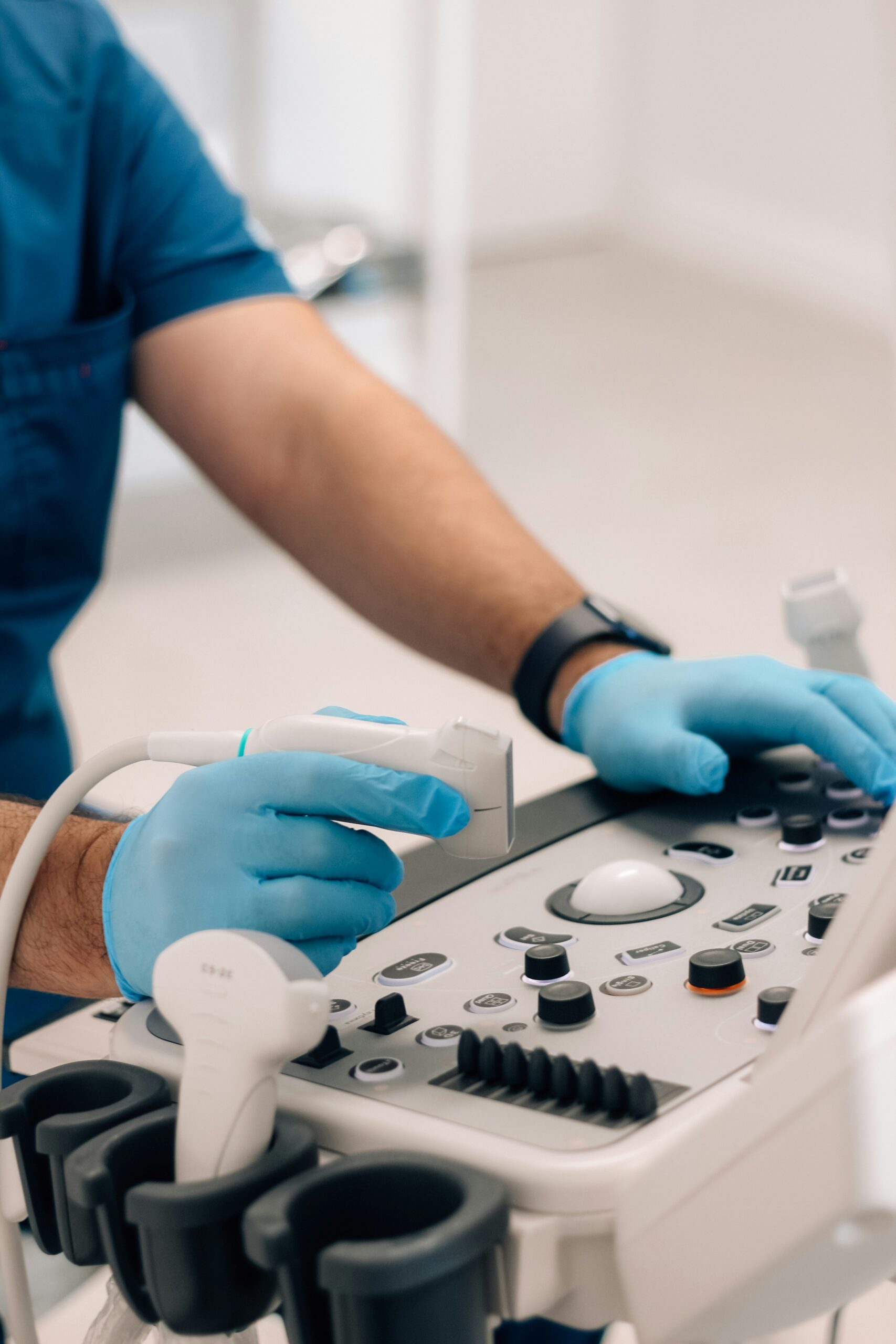
Endometriosis impacts nearly 10 percent of women of reproductive age, often bringing debilitating pain and fertility struggles long before a formal diagnosis is reached. Ultrasound for endometriosis detection offers a non-invasive, readily available window into pelvic anatomy, allowing for the early identification of key lesions like ovarian endometriomas and deep infiltrating implants. In this comprehensive guide, we delve into what endometriosis is, why prompt diagnosis is so vital, ultrasound’s diagnostic precision, procedural protocols (from transvaginal scans to specialized bowel-prepared imaging), and how expert care at a dedicated center elevates patient outcomes. You’ll gain insights into preparing for your scan, understanding your results, and navigating the next steps toward effective treatment.
Understanding Endometriosis and the Importance of Early Diagnosis
Endometriosis is a chronic gynecological condition where tissue similar to the uterine lining grows outside the uterus, triggering inflammation, adhesions, and cyclical bleeding. Early diagnosis is key to preventing progressive damage, alleviating chronic pain, preserving fertility, and guiding timely treatment plans—such as medical therapy or surgical excision—which can significantly enhance your quality of life.
Common Symptoms and the Impact of Endometriosis
Before imaging, patients frequently report:
- Persistent pelvic pain that worsens around menstruation and may extend to the back or legs.
- Severe menstrual cramps (dysmenorrhea) often accompanied by nausea and gastrointestinal distress.
- Pain during intercourse (dyspareunia) impacting intimacy and emotional well-being.
- Infertility, with 30–50 percent of women experiencing conception challenges due to adhesions or ovarian lesions.
These symptoms can lead to fatigue, anxiety, and a diminished capacity for daily activities, making early detection absolutely critical.
Why Diagnostic Delays Occur and Their Impact on Patients
Diagnostic delays, averaging 7–10 years, often stem from the normalization of symptoms, overlap with other gynecological conditions, and limited access to specialized imaging. Each year of delay can lead to lesion progression, intensified pain, and increased complexity in surgical management.
How Ultrasound Aids in the Endometriosis Diagnostic Journey
Ultrasound serves as a primary imaging tool, offering real-time visualization of pelvic structures without the use of ionizing radiation. It effectively detects suggestive signs, particularly endometriomas and deep infiltrating endometriosis, guides further imaging or laparoscopy, and helps categorize disease severity for personalized care plans.
Assessing the Accuracy of Ultrasound in Endometriosis Diagnosis
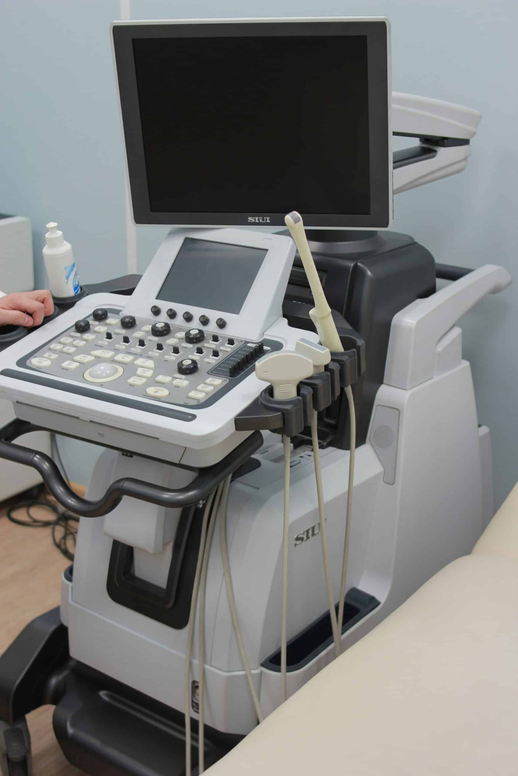
Ultrasound demonstrates high diagnostic performance for moderate to severe endometriosis, achieving a sensitivity of up to 88 percent and a specificity of nearly 79 percent for deep lesions.
Its non-invasive nature and widespread availability make it an ideal initial triage tool in the diagnostic pathway.
Types of Endometriosis Detectable by Ultrasound
Ultrasound is particularly adept at identifying:
- Ovarian Endometriomas – These are cystic “chocolate” lesions characterized by homogeneous, low-level echoes.
- Deep Infiltrating Endometriosis (DIE) – Nodular implants that invade ligaments, the bladder, or the bowel wall.
- Adenomyosis – Diffuse thickening of the uterine wall accompanied by myometrial cysts.
It’s important to note that superficial peritoneal lesions can remain challenging to visualize.
Limitations of Ultrasound in Endometriosis Diagnosis
Key constraints include:
- Limited ability to detect small, superficial peritoneal implants.
- A degree of operator dependence, with variable expertise across different centers.
- A restricted field of view for extra-pelvic disease, such as endometriosis affecting the diaphragm or surgical scars.
These limitations may necessitate the use of complementary imaging techniques.
Comparing Ultrasound to Other Diagnostic Methods: MRI and Laparoscopy
The following comparison highlights the relative strengths of each modality:
Ultrasound is ideal for initial evaluation, MRI helps refine mapping, and laparoscopy provides definitive confirmation.
Understanding Transvaginal Ultrasound for Endometriosis Diagnosis

Transvaginal ultrasound (TVS) utilizes a high-frequency probe inserted into the vagina to generate detailed images of the uterus, ovaries, and surrounding pelvic structures. This method significantly enhances the detection of subtle endometriotic lesions compared to transabdominal scans.
The Transvaginal Ultrasound Procedure for Endometriosis
A TVS procedure typically involves:
- Patient positioning in the lithotomy position with a comfortably full bladder.
- Probe preparation with a protective cover and lubricant for enhanced comfort.
- Systematic scanning of the ovaries, uterosacral ligaments, bladder base, and pouch of Douglas.
- Documentation of any identified cysts, nodules, or adhesions, including precise measurements.
Clear communication and a gentle technique are paramount to ensuring patient comfort and achieving optimal imaging quality.
Benefits of Specialized Endometriosis Ultrasound Protocols (e.g., IDEA Consensus)
Specialized protocols are designed to standardize lesion assessment and reporting:
- The IDEA Consensus provides defined sonographic signs and systematic examination steps.
- High-resolution transducers improve the detection of subtle nodules.
- Bowel preparation protocols minimize artifacts and enhance visualization of posterior deep infiltrating endometriosis.
These measures collectively boost diagnostic confidence and improve inter-observer consistency.
How Transvaginal Ultrasound Detects Endometriomas and Deep Infiltrating Endometriosis
Endometriomas typically appear as unilocular cysts containing homogeneous, low-level internal echoes, often described as having a “ground-glass” texture.
Deep infiltrating endometriosis (DIE) manifests as hypoechoic nodules that tether organs, cause thickening of ligaments, or lead to local tissue retraction. Color Doppler imaging may reveal peripheral vascularity around active implants.
Deep Infiltrating Endometriosis (DIE) and Its Ultrasound Diagnosis
Deep infiltrating endometriosis (DIE) refers to lesions that penetrate more than 5 mm beneath the peritoneal surface, frequently affecting the uterosacral ligaments, rectovaginal septum, bladder, or bowel.
Ultrasound Indicators of Deep Infiltrating Endometriosis (DIE)
Ultrasound markers suggestive of DIE include:
- Hypoechoic nodules observed along ligaments or organ surfaces.
- Tissue retraction leading to organ immobility.
- Uterosacral ligaments that are thickened, exceeding 4 mm in width.
- Bowel wall irregularities indicating localized invasion.
These findings are crucial for precise lesion mapping.
The Importance of Bowel Preparation for DIE Ultrasound Scans
Clearing fecal residues and gas through mild laxatives or enemas:
- Reduces acoustic shadowing caused by gas.
- Enhances the contrast between the bowel wall and adjacent lesions.
- Improves visualization of posterior compartment implants near the rectum.
Optimized imaging leads to more reliable staging of the disease.
Preparing for Your Endometriosis Ultrasound
Thorough preparation is essential to maximize scan quality and diagnostic yield, fostering patient confidence and minimizing the need for repeat examinations.
Recommended Bowel Preparation for Specialized Endometriosis Ultrasound
A typical bowel preparation protocol may involve:
- A low-residue diet for 24 hours preceding the scan.
- A mild oral laxative taken the evening before the appointment.
- A tap water enema administered two hours prior to imaging to ensure clear rectal visualization.
Adhering to these steps helps reduce artifacts and ensures clear delineation of lesions.
What to Expect During Your Ultrasound Procedure
During the scan, you can expect:
- Comfortable positioning with options for privacy and support.
- Minimal discomfort as the transvaginal probe is gently maneuvered.
- Real-time feedback on detected findings, promoting transparency throughout the process.
Technologists and physicians work collaboratively to ensure a reassuring and supportive environment.
Understanding and Interpreting Your Ultrasound Results
Following your imaging session, you will receive:
- Structured reports detailing lesion location, size, and suspected depth.
- Annotated images highlighting key findings for enhanced clarity.
- Expert review by a gynecologist or radiologist who will explain the implications and outline the next steps.
Clear explanations empower you to participate actively in shared decision-making regarding your care.
Next Steps Following an Endometriosis Ultrasound Diagnosis
The findings from your ultrasound will guide a personalized diagnostic and treatment journey, ensuring you proceed along the most appropriate pathway.
When MRI is Recommended After Ultrasound
An MRI may be indicated when:
- Complex DIE is suspected across multiple pelvic compartments.
- Deep bladder or ureteric involvement requires higher soft-tissue contrast for detailed assessment.
- Ultrasound findings are inconclusive for mapping multifocal disease.
MRI complements ultrasound by providing refined details crucial for surgical planning.
Why Laparoscopy is Considered the Gold Standard for Diagnosis
Laparoscopy offers:
- Direct visualization of all lesion types, including subtle superficial peritoneal implants.
- Histological confirmation through biopsy, ensuring a definitive diagnosis.
- Simultaneous treatment via excision or ablation during the same procedure.
It remains the conclusive diagnostic and therapeutic approach for endometriosis.
How Ultrasound Findings Influence Treatment Options
Sonographic staging plays a key role in determining:
- Medical management, such as hormonal suppression for minimal to mild disease.
- Conservative excision of endometriomas to preserve ovarian reserve.
- Extensive surgical intervention for multifocal DIE, often requiring multidisciplinary teams.
Imaging-driven personalization enhances both the efficacy and safety of treatment.
The Critical Importance of Expert Ultrasound Diagnosis at a Specialized Center
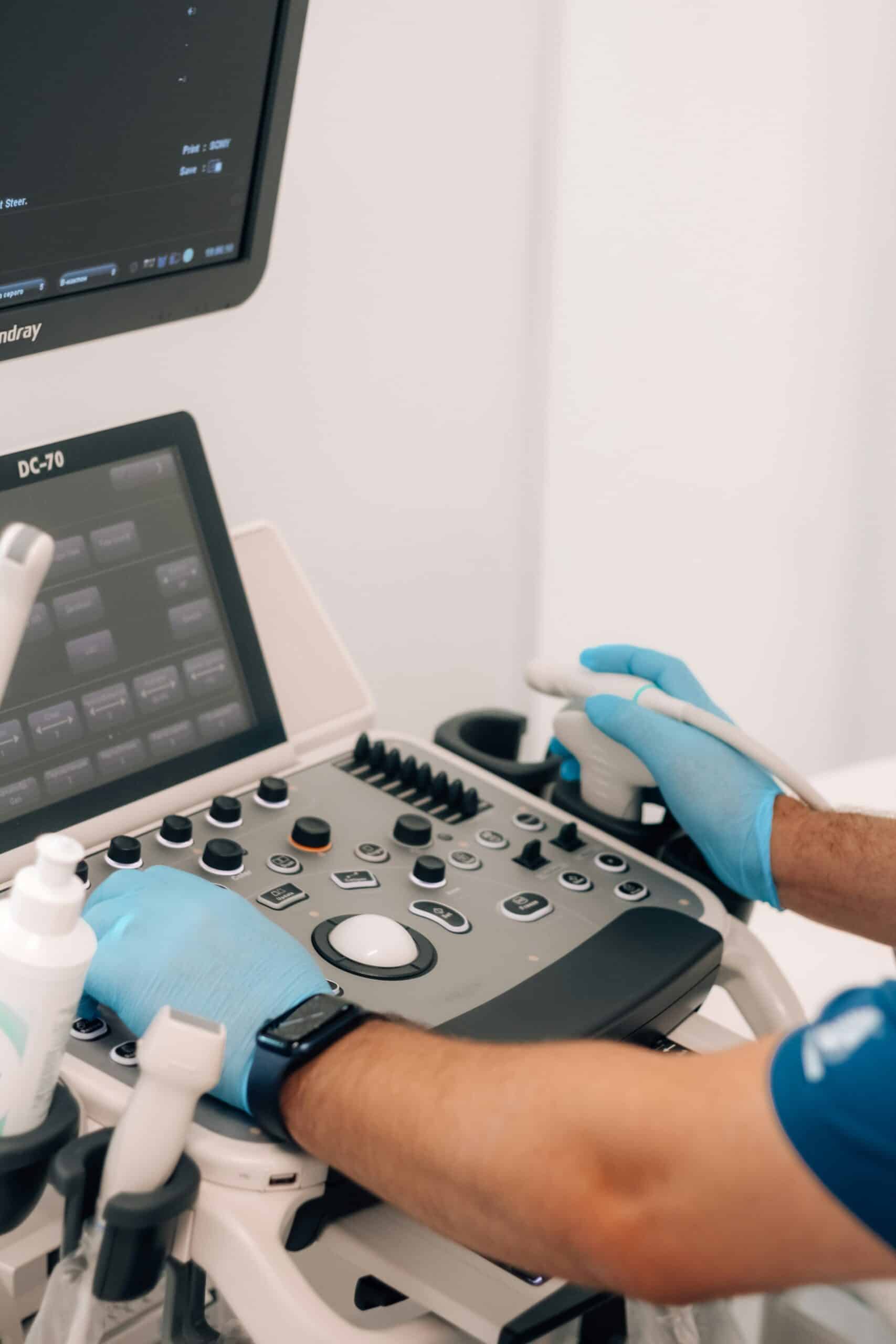
Accessing a dedicated endometriosis center ensures that your imaging, interpretation, and subsequent care benefit from concentrated expertise and advanced technology.
How Expertise Enhances Ultrasound Accuracy and Patient Outcomes
Specialized sonographers and physicians:
- Adhere to consensus protocols for consistent and accurate lesion detection.
- Achieve higher inter-observer agreement through advanced training and experience.
- Translate imaging findings into individualized treatment strategies designed to minimize complications.
This specialized expertise reduces the likelihood of missed diagnoses and optimizes therapeutic success.
Advanced Imaging Technologies at Specialized Centers
Dedicated centers often utilize:
- High-frequency transducers for superior spatial resolution and finer detail.
- Three-dimensional ultrasound reconstructions for volumetric assessment of lesions.
- Elastography to help differentiate fibrotic nodules from benign tissue.
These technological advancements deepen diagnostic insight and precision.
Enhancing the Diagnostic Experience with a Patient-Centered Approach
A center of excellence prioritizes:
- Comprehensive consultations that clearly explain the rationale behind imaging and the findings.
- Multidisciplinary collaboration involving gynecology, radiology, and pain management specialists.
- Emotional support through counseling services and peer support networks.
This compassionate framework transforms a clinical procedure into an empowering step toward recovery and well-being.
Frequently Asked Questions About Ultrasound and Endometriosis Diagnosis
Can Ultrasound Definitive Diagnose Endometriosis?
Ultrasound is highly effective at identifying characteristic lesions like endometriomas and deep infiltrating implants, but it cannot visualize every superficial peritoneal nodule. While it offers high reliability for moderate to severe disease, a normal ultrasound result does not entirely exclude endometriosis, and laparoscopy remains the definitive diagnostic standard.
What Does Endometriosis Appear As on Ultrasound?
On ultrasound, endometriotic changes typically present as:
- Homogeneous, low-level echoes within ovarian cysts, often described as a “ground-glass” appearance in endometriomas.
- Hypoechoic nodules or thickening of ligaments, indicative of DIE.
- Myometrial cysts or a heterogeneous uterine wall, consistent with adenomyosis.
Is It Possible to Have Endometriosis with a Normal Ultrasound?
Yes. Small superficial implants and early-stage disease may not be detected on either transvaginal or transabdominal scans. This underscores the importance of clinical correlation and, if necessary, further evaluation with MRI or laparoscopy.
How Accurate Is Ultrasound for Detecting Deep Infiltrating Endometriosis?
When performed by experienced operators utilizing consensus guidelines, ultrasound demonstrates a sensitivity of up to 88 percent and a specificity of nearly 79 percent for DIE. Bowel preparation and the use of high-resolution probes significantly contribute to these accuracy rates.
Is Bowel Preparation Always Necessary for Endometriosis Ultrasound?
Bowel preparation is considered essential for evaluating the posterior compartment and DIE. However, it may be optional when the primary focus is on assessing ovarian endometriomas or adenomyosis. A tailored approach balances patient convenience with the need for diagnostic clarity.
Endometriosis diagnosis through ultrasound represents a pivotal, non-invasive step toward timely treatment and symptom relief. Expert-led imaging at a specialized center not only enhances detection accuracy but also fosters a patient-centered journey through diagnosis, planning, and care. For women and families navigating endometriosis, advanced ultrasound protocols provide clarity, reduce uncertainty, and empower informed decisions about treatment pathways.
Conclusion
Ultrasound has become an indispensable first step in identifying endometriosis, offering a non-invasive way to detect endometriomas, deep infiltrating lesions, and adenomyosis with strong diagnostic accuracy. While it cannot replace laparoscopy as the definitive diagnostic standard, it plays a central role in triaging patients, guiding further imaging such as MRI, and shaping personalized treatment plans, from medical management to surgical excision.
Careful preparation, including bowel protocols for posterior compartment evaluation, and interpretation by experienced sonographers and physicians following consensus guidelines, can make a meaningful difference in diagnostic accuracy. Choosing a specialized center for this imaging ensures patients benefit from advanced technology, multidisciplinary expertise, and compassionate, patient-centered care throughout the diagnostic journey. With timely, expert-led ultrasound evaluation, patients living with endometriosis symptoms can move forward with clarity and a clear path toward effective treatment.