
Pinpointing endometriosis accurately can significantly shorten the typical seven- to ten-year diagnostic delay by employing targeted tests. We’ll explore how these tests balance invasiveness, cost, and detection rates, covering laparoscopy, ultrasound, MRI, and promising new non-invasive options. This comparison delves into endometriosis pathophysiology and symptom triggers, explains why laparoscopy remains the gold standard, evaluates imaging modalities, highlights promising blood and saliva biomarkers, and offers a head-to-head analysis of accuracy, recovery time, and expenses. Along the way, we’ll address diagnostic challenges, cost considerations, and guide you toward specialized expert care for a personalized diagnostic journey.
What Is Endometriosis and Why Is Accurate Diagnosis Important?

Endometriosis occurs when tissue similar to the uterine lining grows outside the uterus, triggering chronic inflammation, pain, and fertility issues. Diagnosing it precisely ensures timely treatment and helps prevent long-term complications. This section defines the key symptoms and patient impacts that prompt testing, setting the stage for comparing diagnostic methods.
What Are the Common Symptoms Prompting Endometriosis Testing?
Endometriosis most often presents as pelvic pain, painful periods, and discomfort during intercourse, prompting women to seek medical evaluation.
-
Persistent pelvic pain that extends beyond menstrual cycles.
-
Severe menstrual cramps that interfere with daily life.
-
Pain during sexual intercourse, particularly with deep penetration.
-
Difficulty conceiving after one year of trying without contraception.
These symptoms frequently overlap with other conditions, leading patients down a complex diagnostic path and highlighting the need for specialized evaluation.
How Does Endometriosis Impact Women’s Health and Fertility?
Endometriosis causes inflammation and scarring around reproductive organs, potentially reducing ovarian reserve, distorting pelvic anatomy, and creating adhesions that hinder egg release or embryo implantation. Inflammatory substances released by these growths can also affect embryo development, making infertility a common concern. Addressing these issues early can help preserve fertility and manage chronic pain, guiding clinicians toward the most effective diagnostic approach.
Why Is Early and Precise Diagnosis Critical?
Early and accurate identification of endometriosis can prevent lesion progression, protect ovarian function, and optimize pain management through tailored hormonal or surgical treatments. For instance, promptly identifying deep infiltrating lesions can prevent complications involving the bowel or bladder and preserve a patient’s quality of life. Establishing the presence and extent of the disease from the outset ensures patients receive the correct treatment and reduces the need for repeat procedures.
Why Is Laparoscopy Considered the Gold Standard for Endometriosis Diagnosis?

Laparoscopy is a minimally invasive surgical procedure that allows direct visualization of endometrial implants and enables tissue biopsy for histological confirmation, providing a definitive diagnosis.
What Happens During a Laparoscopy Procedure?
During laparoscopy, a thin, lighted tube called a laparoscope is inserted through a small incision in the abdomen, typically under general anesthesia, to examine the pelvic cavity for signs of endometriosis. Carbon dioxide gas is used to create space for better visualization, and small instruments are used to remove suspicious implants or collect tissue samples for biopsy. This direct view, combined with microscopic examination of the tissue, offers the highest diagnostic accuracy and may allow for simultaneous removal of the lesions.
How Does Histological Confirmation via Biopsy Support Diagnosis?
Biopsy samples taken from suspected endometriotic areas are examined under a microscope to identify endometrial glands and tissue outside the uterus, confirming active disease. Histological evidence not only validates what is seen during surgery but can also indicate the severity of the lesions, which helps in planning postoperative hormone therapy. This level of certainty is unmatched by imaging or biomarkers alone and significantly influences the subsequent treatment plan.
What Are the Benefits and Risks of Laparoscopy?
Laparoscopy offers definitive confirmation, detailed mapping of lesions, and the potential for immediate surgical removal, which can alleviate pain and preserve fertility, though it does carry surgical risks.
-
BenefitsGold-standard accuracy through direct visualization and biopsy.Simultaneous surgical removal reduces the extent of disease.Detailed staging informs personalized treatment plans.
-
RisksComplications related to anesthesia, such as nausea or breathing issues.Potential for bleeding, infection, or injury to nearby organs.Postoperative pain and a recovery period of 1–2 weeks.
Weighing these factors carefully requires a thorough discussion with a specialized gynecologic surgeon to ensure the best possible outcomes.
How Does Laparoscopy Compare to Other Diagnostic Methods?
Laparoscopy boasts over 95% sensitivity and specificity, surpassing imaging techniques whose detection rates can vary significantly for superficial versus deep lesions. In contrast, transvaginal ultrasound might miss peritoneal implants, and MRI can sometimes underdetect small endometriomas. While more invasive and costly, laparoscopy remains the only method to confirm the diagnosis through histology, setting the benchmark for all non-surgical tests.
How Do Ultrasound and MRI Imaging Tests Compare for Endometriosis Detection?
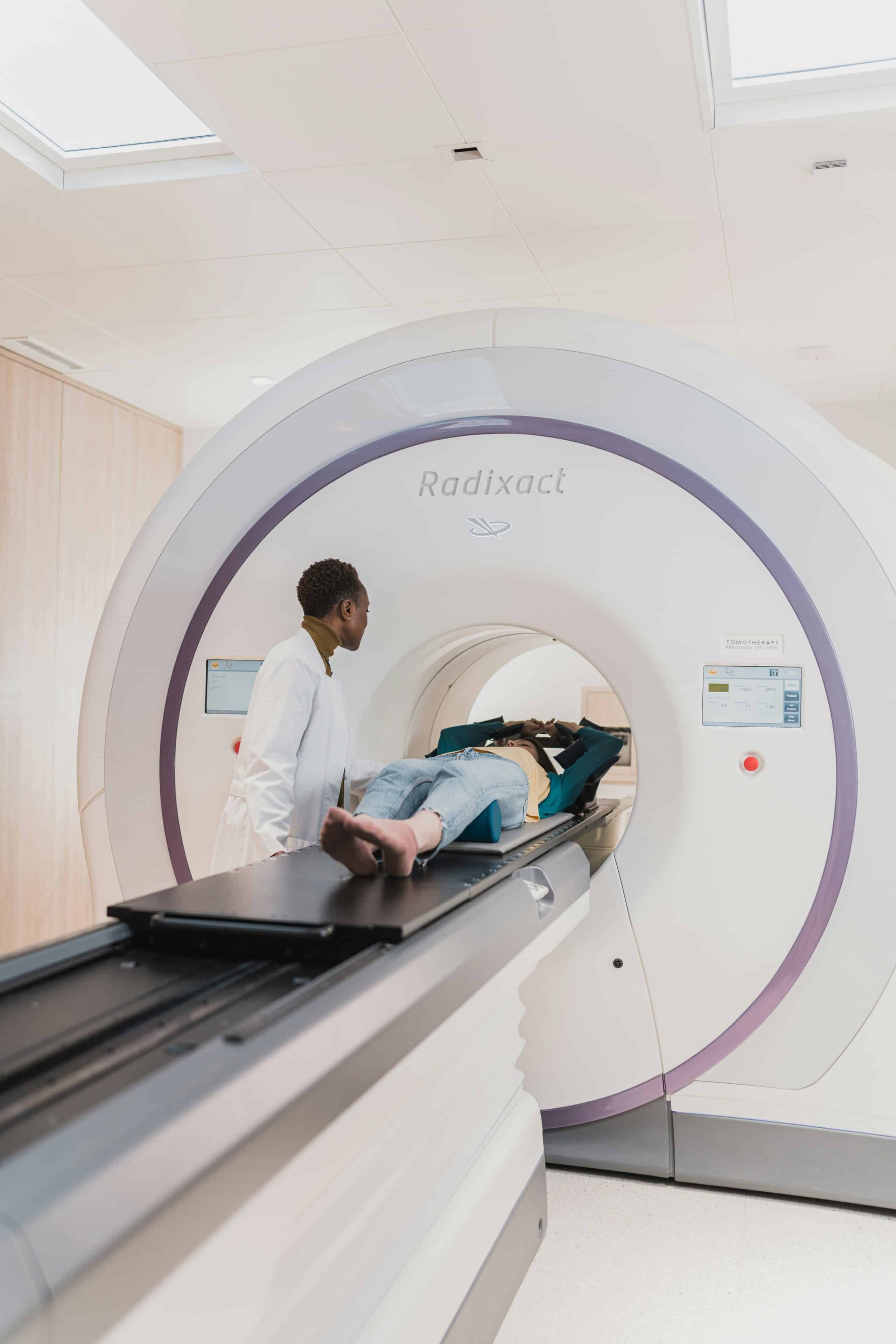
Non-invasive imaging techniques provide detailed views of ovarian cysts and deep infiltrating lesions, aiding in surgical planning. However, each method has its own strengths and limitations. The following overview clarifies when to choose ultrasound versus MRI, and how cost factors into these decisions.
What Can Transvaginal Ultrasound Detect in Endometriosis?
Transvaginal ultrasound uses high-frequency sound waves transmitted via a vaginal probe to identify ovarian endometriomas (chocolate cysts), nodules in the uterosacral ligaments, and involvement of the rectovaginal septum. Its real-time imaging is particularly effective for assessing endometriomas larger than 1 cm and guiding procedures like aspiration or drainage.
What Are the Strengths and Limitations of Ultrasound?
Transvaginal ultrasound is a cost-effective, widely available option that doesn’t involve radiation. However, it can struggle to detect superficial peritoneal lesions and small implants located near the bowel.
-
StrengthsHigh accuracy (over 90%) for detecting ovarian endometriomas.Dynamic assessment with Doppler technology to evaluate blood flow in lesions.Lower cost, minimal preparation required, and immediate results.
-
LimitationsAccuracy can depend heavily on the skill of the operator and requires extensive training.Limited view of deep pelvic areas.Not ideal for mapping superficial peritoneal disease.
This variability underscores the importance of using ultrasound in conjunction with other techniques when precise staging is necessary.
How Does MRI Enhance Detection of Deep Infiltrating Endometriosis?
Magnetic resonance imaging (MRI) offers high-contrast resolution of soft tissues, enabling the identification of deep infiltrating endometriosis in the bowel, bladder, ureters, and pelvic sidewalls. Specialized MRI protocols can precisely map the depth and extent of lesions, guiding surgeons to areas requiring targeted removal and potentially shortening operative times.
When Should MRI Be Preferred Over Ultrasound?
MRI is the preferred choice when ultrasound results are unclear, when deep infiltrating disease is suspected, or when comprehensive preoperative mapping is crucial. Patients who have had previous surgeries, have complex pelvic anatomy, or experience persistent symptoms despite a normal ultrasound often benefit most from MRI’s broader imaging capabilities.
What Are the Cost Differences Between Ultrasound and MRI?
While costs vary by location and provider, transvaginal ultrasound typically ranges from $200 to $500, whereas a pelvic MRI can cost between $800 and $2,500. Insurance coverage often favors ultrasound as an initial diagnostic step, with MRI typically reserved for surgical planning or when initial imaging is inconclusive. Understanding these out-of-pocket expenses can help patients and providers select the most cost-effective approach without compromising diagnostic accuracy.
What Are Emerging Non-Invasive Tests for Endometriosis Diagnosis?
Blood, saliva, and electrophysiological tests are showing promise in reducing invasiveness and shortening the diagnostic timeline by detecting biomarkers or electrical changes associated with endometrial tissue activity. These investigational tests aim to complement imaging and laparoscopy in earlier screening.
How Reliable Are Blood Tests Like CA-125 and DotEndo?
Blood tests can measure CA-125 levels or analyze novel biomarker panels like DotEndo, which detect inflammatory proteins and extracellular vesicles linked to ectopic endometrial activity. CA-125 has a sensitivity of 50–60% for advanced disease but lacks specificity, as its levels can be elevated in other conditions.
DotEndo has shown up to 85% accuracy in early studies, but larger validation is needed before it can replace invasive diagnostic methods.
What Is the Role of Saliva-Based miRNA Tests in Diagnosis?
Saliva-based microRNA profiling identifies RNA fragments released by endometrial lesions into bodily fluids, offering potential for non-invasive, rapid screening. Early research suggests certain miRNA patterns are associated with the presence of lesions, but standardization and large-scale clinical trials are still underway before these tests can be used routinely.
How Do Electrophysiological Tests Like EndoSure and EndoSearch Work?
Electrophysiological diagnostic tools measure changes in electrical impedance or conductivity within the pelvic region caused by ectopic tissue. Devices like EndoSure create electrical maps to pinpoint lesion locations without the need for imaging. Preliminary data show promise in locating deep implants, although their sensitivity for superficial lesions is still being evaluated.
What Is the Future Potential of Non-Invasive Endometriosis Testing?
Ongoing research into multi-omics panels, point-of-care assays, and artificial intelligence–driven pattern recognition aims to develop a non-invasive diagnostic algorithm with over 90% accuracy. Combining biomarker data with imaging results could reduce reliance on laparoscopy and speed up the initiation of treatment.
How Do Endometriosis Diagnostic Tests Compare in Accuracy, Invasiveness, and Cost?
No single test perfectly balances all diagnostic attributes. The table below compares key metrics to help patients and providers choose the most suitable option based on the type of disease, urgency, and financial considerations.
Each diagnostic method presents trade-offs between sensitivity, specificity, and the patient’s experience. Selecting the ideal approach depends on symptom severity, suspicion of lesions, and available resources, guiding a personalized diagnostic pathway.
What Challenges Affect the Endometriosis Diagnostic Journey?
Numerous factors contribute to prolonged delays and misdiagnoses, impacting patients’ quality of life and complicating treatment planning. Understanding these obstacles can inform strategies to streamline evaluations at specialized centers.
Why Is There an Average 7-10 Year Delay in Diagnosis?
Endometriosis symptoms often mimic common gynecologic and gastrointestinal conditions, leading to pelvic pain being mistakenly attributed to IBS, pelvic inflammatory disease, or ovarian cysts. Limited awareness among primary care providers and the varied ways symptoms can present contribute to extensive referral pathways and delayed specialist consultations. Addressing these gaps through education and direct referral pathways can shorten the diagnostic timeline.
Which Conditions Commonly Mimic Endometriosis Symptoms?
Symptoms such as chronic pelvic pain and painful periods can overlap with:
-
Irritable bowel syndrome (IBS)
-
Interstitial cystitis/painful bladder syndrome
-
Pelvic inflammatory disease (PID)
-
Uterine fibroids
-
Ovarian torsion or cyst rupture
Differential diagnosis through targeted testing ensures that endometriosis is neither over- nor under-diagnosed.
How Does Specialist Expertise Improve Diagnostic Accuracy?
Gynecologic surgeons and radiologists with specialized training in endometriosis are adept at recognizing subtle imaging signs and lesion patterns, ensuring the appropriate tests are selected and interpreted correctly. Centers of excellence utilize multidisciplinary teams for integrated ultrasound, MRI, and surgical assessments, leading to higher detection rates and fewer repeat surgeries.
What Emotional and Practical Challenges Do Patients Face?
Patients often experience chronic pain, anxiety about fertility, and frustration from undergoing multiple inconclusive tests. Financial burdens from numerous consultations and out-of-pocket expenses add to the stress. Comprehensive care teams offering counseling, pain management, and financial guidance can bolster patient resilience and encourage adherence to diagnostic and treatment plans.
When Should You Seek Expert Care and How to Choose the Right Diagnostic Path?
Engaging a specialized endometriosis center early on provides focused evaluation, coordinated multidisciplinary care, and personalized testing strategies that maximize the chances of an accurate diagnosis. Understanding when and how to seek expert consultation can lead to a smoother diagnostic journey.
What Are the Signs You Need a Specialist for Endometriosis Diagnosis?
A referral to a gynecologist with expertise in endometriosis is recommended when:
-
Pelvic pain or pain during intercourse persists despite initial treatments.
-
Infertility remains unexplained after standard fertility assessments.
-
Imaging reveals complex ovarian cysts or suspicious deep lesions.
-
Multiple potential diagnoses have been ruled out without symptom resolution.
Timely referral facilitates targeted investigations and reduces the need for unnecessary procedures.
How Does a Center of Excellence Enhance Diagnostic and Treatment Outcomes?
Specialized centers integrate advanced imaging, surgical expertise, pain management specialists, and reproductive endocrinologists to deliver coordinated care. This team-based approach allows for precise lesion mapping, individualized therapy, and streamlined follow-up, resulting in higher diagnostic accuracy and improved patient satisfaction.
How Can Personalized Diagnostic Approaches Improve Accuracy?
Tailoring test selection to a patient’s specific symptoms and suspected lesion type—such as combining transvaginal ultrasound with targeted MRI for suspected rectovaginal disease—can enhance detection rates. Personalized biomarker panels and electrophysiological mapping further refine the diagnostic process, minimizing the need for invasive procedures whenever possible.
How to Navigate Costs and Insurance for Endometriosis Diagnosis?
Understanding insurance coverage for imaging and surgery, obtaining preauthorization, and exploring financial assistance programs can significantly reduce out-of-pocket expenses. Patient advocates at specialized centers can often guide patients toward cost-effective options, ensuring that financial barriers do not delay essential diagnostic testing.
What Resources and Support Are Available During the Diagnostic Process?
Support services include educational materials, peer support groups, counseling for chronic pain, fertility guidance, and information on research trials. Accessing these resources empowers patients with knowledge and community as they navigate complex diagnostic pathways.
Frequently Asked Questions
What is the gold-standard test for diagnosing endometriosis?
Laparoscopy combined with histological biopsy remains the gold standard, offering direct visualization of implants and microscopic confirmation of disease with over 95% sensitivity and specificity. It is the only method that provides definitive histological proof, though it is a surgical procedure that carries recovery time and surgical risk.
Can endometriosis be diagnosed without surgery?
Transvaginal ultrasound and pelvic MRI can identify ovarian endometriomas and deep infiltrating lesions non-invasively, and emerging blood tests such as CA-125 and the DotEndo panel are being studied as screening tools. These non-invasive options can guide suspicion and surgical planning, but none yet matches the diagnostic certainty of laparoscopic biopsy.
Why does it typically take seven to ten years to diagnose endometriosis?
Endometriosis symptoms often mimic conditions like irritable bowel syndrome, interstitial cystitis, and pelvic inflammatory disease, which leads to misattributed pain and delayed referrals. Limited awareness among primary care providers and the absence of a simple, routine screening test further extend the diagnostic timeline.
How do ultrasound and MRI differ in detecting endometriosis?
Transvaginal ultrasound is a lower-cost, widely available first step that performs well for ovarian endometriomas but can miss superficial peritoneal implants. MRI offers superior soft-tissue contrast for mapping deep infiltrating disease in the bowel, bladder, or pelvic sidewalls, making it the preferred choice when ultrasound findings are unclear or deep disease is suspected.
When should someone seek a specialist for endometriosis testing?
A referral to a gynecologist with endometriosis expertise is warranted when pelvic pain or pain during intercourse persists despite treatment, infertility remains unexplained, imaging shows complex ovarian cysts or suspicious deep lesions, or other diagnoses have been ruled out without symptom relief. Specialized centers coordinate imaging, surgical, and biomarker testing to reach an accurate diagnosis faster.
Conclusion
Endometriosis diagnosis spans a spectrum of tools, from cost-effective imaging like transvaginal ultrasound to the surgical certainty of laparoscopy with histological biopsy, each offering a distinct balance of accuracy, invasiveness, and expense. Emerging blood, saliva, and electrophysiological tests hold promise for earlier, less invasive screening, but they currently complement rather than replace established methods. Because symptoms so often mimic other conditions, the path to an accurate diagnosis depends on recognizing warning signs early and seeking evaluation at a specialized center equipped to coordinate imaging, surgical expertise, and personalized testing. With the right diagnostic approach, women can move past years of uncertainty toward timely, effective treatment that protects both fertility and quality of life.