

A diagnosis of deep infiltrating endometriosis may sound like one specific condition, but the disease does not necessarily look or behave the same way in every patient. One person may have endometriosis affecting tissue behind the uterus, while another may have suspected involvement near the bowel, bladder, or ureters.
These differences may influence the symptoms a patient notices, the imaging that could be useful, the specialists who may need to participate in care, and the questions that should be discussed before treatment.
Location is only one part of the picture. Symptoms, previous treatments, fertility priorities, organ function, imaging findings, and personal preferences may also contribute to the care plan. Understanding how these factors fit together can help patients have clearer, more productive conversations with an endometriosis specialist.
One Diagnosis Can Describe Several Disease Patterns

Deep infiltrating endometriosis generally refers to endometriosis that extends beneath the surface of affected tissue. It may develop in different areas of the pelvis, including the tissue behind the uterus, supporting ligaments, the space between the vagina and rectum, the bowel, the bladder, or the tissue surrounding the ureters.
Not every patient will have disease in all of these areas. Even when two patients have lesions in a similar location, their symptoms and treatment priorities may differ. This is why a diagnosis alone may not provide enough information to determine the next step.
Disease Behind the Uterus and Along Supporting Ligaments
The uterosacral ligaments are bands of tissue that help support the uterus and connect it with structures toward the back of the pelvis. Deep endometriosis may develop along these ligaments or in nearby tissue behind the uterus.
Patients with disease in this region may report deep pelvic pain, discomfort during or after intercourse, painful periods, lower back discomfort, or a feeling of pressure in the pelvis. However, these symptoms can have several possible causes, so they cannot confirm the location of endometriosis by themselves.
Endometriosis may also contribute to inflammation, scar tissue, or adhesions that limit the normal movement of pelvic structures. A patient may therefore experience pain during activities that place pressure on the pelvis, even when the discomfort is not limited to menstruation.
A detailed symptom history, pelvic examination when appropriate, and review of relevant imaging may help a specialist decide whether this area requires closer evaluation.
Rectovaginal and Vaginal Involvement
The rectovaginal area is the tissue between the vagina and rectum. Endometriosis in or near this space may be associated with deep pain during intercourse, pain with bowel movements, rectal pressure, pelvic discomfort, or symptoms that become more noticeable around menstruation.
These symptoms do not always mean that endometriosis has entered the bowel wall or vagina. Disease in nearby tissue, pelvic floor muscle tension, adhesions, and other pelvic pain conditions may create similar experiences.
The purpose of specialist evaluation is therefore not simply to match one symptom with one organ. It is to understand the overall pattern and determine which structures may be contributing to pain or functional concerns.
Disease Near the Pelvic Sidewall
The pelvic sidewall contains important structures, including blood vessels, nerves, connective tissue, and the ureters, which carry urine from the kidneys to the bladder.
Endometriosis in this region may require careful evaluation because several structures are located close together. Pain may sometimes be felt in the pelvis, hip, back, groin, or upper leg, although these symptoms can also arise from musculoskeletal, nerve, or pelvic floor conditions.
When sidewall involvement is suspected, the care team may review imaging and previous surgical records closely. The goal is to understand the anatomy before treatment is recommended rather than assuming that every area of pain represents a visible endometriosis lesion.
Bowel and Urinary Tract Involvement Raise Different Questions

Deep infiltrating endometriosis may affect structures outside the reproductive organs. Bowel, bladder, and ureteral involvement are often discussed together, but each area may present different symptoms and treatment considerations.
Current guidance recommends specialist referral when deep endometriosis involving the bowel, bladder, or ureter is suspected or confirmed. Evaluation may include targeted ultrasound, MRI in selected circumstances, and input from other specialists when appropriate.
Rectal and Sigmoid Colon Involvement
The rectum and sigmoid colon are located close to the uterus, vagina, and other pelvic structures. Deep endometriosis may develop on the outer surface of the bowel or extend into deeper layers.
Possible symptoms may include:
-
Pain during bowel movements
-
Constipation, diarrhea, or alternating bowel habits
-
Rectal pressure
-
Bloating
-
A feeling of incomplete emptying
-
Bowel symptoms that change around menstruation
Pain during bowel movements
Constipation, diarrhea, or alternating bowel habits
Rectal pressure
Bloating
A feeling of incomplete emptying
Bowel symptoms that change around menstruation
These concerns are not specific to endometriosis. Irritable bowel syndrome, pelvic floor dysfunction, food intolerances, inflammatory conditions, and other digestive disorders may produce overlapping symptoms.
When bowel endometriosis is being considered, the care team may look at more than symptom severity. The location of a suspected lesion, its relationship to the bowel wall, possible narrowing, adhesions, prior surgery, and the patient’s overall bowel function may all be relevant.
A bowel lesion seen on imaging does not automatically determine the treatment. Some patients may be candidates for medical symptom management or observation, while others may discuss surgery because of pain, functional concerns, fertility planning, or the suspected extent of disease. The decision should be individualized.
Bladder Involvement Versus Bladder-Like Symptoms
Urinary urgency, frequent urination, pelvic pressure, discomfort as the bladder fills, or pain during urination may occur in patients with endometriosis. However, these symptoms do not necessarily mean that an endometriosis lesion is growing within the bladder wall.
Similar symptoms may be associated with:
-
Pelvic floor muscle tension
-
Bladder pain syndrome
-
Recurrent urinary infections
-
Irritation around the bladder
-
Adhesions
-
Endometriosis near, rather than within, the bladder
-
Other gynecologic or urologic conditions
Pelvic floor muscle tension
Bladder pain syndrome
Recurrent urinary infections
Irritation around the bladder
Adhesions
Endometriosis near, rather than within, the bladder
Other gynecologic or urologic conditions
The timing of symptoms may provide useful context. For example, urinary discomfort that repeatedly changes around the menstrual cycle may prompt further endometriosis evaluation. Still, timing alone cannot confirm where the symptoms originate.
A specialist may consider the patient’s history, urine testing, examination findings, imaging, and whether a urologic evaluation would be useful. This broader approach can help avoid assuming that all urinary symptoms have one cause.
Why Ureteral Involvement Is Considered Separately
The ureters are narrow tubes that transport urine from the kidneys to the bladder. Endometriosis may sometimes affect the tissue around a ureter or, less commonly, involve deeper portions of the ureter itself.
Some patients may experience pelvic, back, or urinary symptoms, while others may have few noticeable signs. Understanding the silent kidney risks of ureteral endometriosis is important because symptoms may not always reflect the degree of involvement.
This does not mean that every patient with deep endometriosis requires extensive urinary testing. The need for further evaluation depends on the suspected disease location, imaging findings, symptoms, medical history, and specialist judgment.
When meaningful ureteral involvement is anticipated, coordination with a urologist may be considered before surgery. Advance planning may allow the care team to discuss possible procedures and organ-preservation goals with the patient rather than making unexpected decisions during an operation.
Symptoms and Imaging Provide Different Pieces of Information
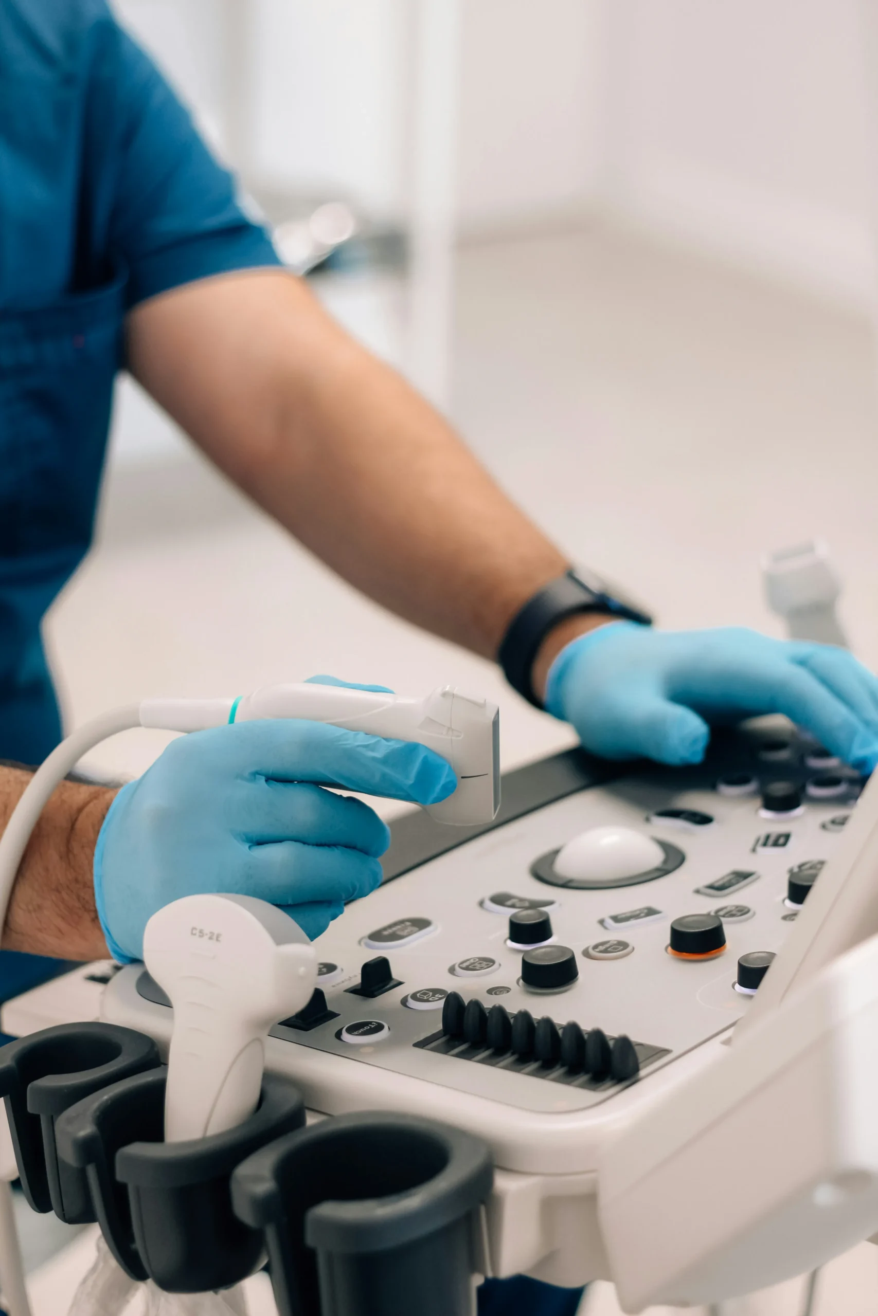
Symptoms and imaging can both contribute to an evaluation, but they answer different questions. A symptom history describes what the patient is experiencing and how those symptoms affect daily life. Imaging may provide information about pelvic anatomy, visible lesions, adhesions, endometriomas, or possible involvement of certain organs. Neither source of information should necessarily be interpreted in isolation.
Symptom Patterns May Suggest an Area but Cannot Confirm It
Certain patterns may encourage a specialist to examine a particular part of the pelvis more closely. For example:
-
Deep pain during intercourse may prompt evaluation of structures behind the uterus, the pelvic floor, or the rectovaginal area.
-
Painful bowel movements that worsen around menstruation may raise concern for posterior pelvic or bowel involvement.
-
Cyclical urinary discomfort may lead to closer assessment of the bladder and urinary tract.
-
Pain radiating into the hip, back, or leg may encourage evaluation of the pelvic sidewall, muscles, nerves, and other possible pain sources.
Deep pain during intercourse may prompt evaluation of structures behind the uterus, the pelvic floor, or the rectovaginal area. You may also find Deep Ligament Pain: Why Sitting, Walking & Intercourse Hurt helpful.
Painful bowel movements that worsen around menstruation may raise concern for posterior pelvic or bowel involvement.
Cyclical urinary discomfort may lead to closer assessment of the bladder and urinary tract.
Pain radiating into the hip, back, or leg may encourage evaluation of the pelvic sidewall, muscles, nerves, and other possible pain sources.
These patterns provide clues rather than a diagnosis. A patient may have severe pain without extensive visible disease, while another may have deeper anatomical involvement with less obvious symptoms.
Pain can also continue after the original source of irritation has changed. Muscles may tighten in response to long-term discomfort, nerves may become more sensitive, and the body may develop protective movement patterns. For this reason, locating endometriosis lesions may be only one part of understanding persistent pelvic pain. We cover this further in Endometriosis Pain: Incomplete Removal vs. True Recurrence.
What Specialist Ultrasound and MRI May Add
Transvaginal ultrasound is commonly used during endometriosis evaluation. When performed and interpreted by clinicians familiar with the condition, it may help identify ovarian endometriomas and signs of deep disease involving structures such as the bowel, bladder, or ureters.
Pelvic MRI may be considered when more detailed anatomical mapping could influence treatment or surgical planning. It may provide information about the location of suspected lesions, relationships between pelvic organs, and areas that may need closer attention.
Guidance from the National Institute for Health and Care Excellence recommends transvaginal ultrasound during the evaluation of suspected endometriosis, including when a pelvic or abdominal examination is normal. It also notes that pelvic MRI may be considered before operative laparoscopy for deep disease involving the bowel, bladder, or ureter.
Imaging is most useful when interpreted alongside the patient’s symptoms, examination findings, previous treatment, and goals. A scan should not be expected to answer every question or decide the care plan by itself.
A Normal Scan Does Not Necessarily End the Evaluation
A normal ultrasound or MRI does not always exclude endometriosis. Some lesions may be too small, superficial, or difficult to distinguish on imaging. The visibility of disease can also depend on the imaging technique, the area involved, and the experience of the person performing or interpreting the study.
NICE guidance specifically advises clinicians not to rule out endometriosis solely because an examination or ultrasound appears normal. Read more in The Importance of Couples Counseling for Endometriosis Challenges.
When symptoms continue, the next step may involve reviewing the diagnosis, considering other possible causes, obtaining specialist imaging, trying medical symptom management, or discussing whether surgical evaluation is appropriate. The most suitable path depends on the individual patient rather than one test result. Read more in The Evolution of Endometriosis: From Ancient to Modern Times.
The Care Plan Should Not Be Based on a Scan Alone

Imaging may help describe anatomy, but it does not automatically show how much a patient is suffering, which symptoms are caused by endometriosis, or which treatment is most appropriate.
A treatment decision may involve balancing several considerations, including pain, bowel or urinary function, fertility goals, previous treatments, surgical risks, other health conditions, and the patient’s preferences.
A Positive Scan Does Not Automatically Mean Surgery
Finding suspected deep endometriosis on imaging does not necessarily mean that an operation is required immediately. Some patients may prefer to begin or continue medical symptom management, pelvic floor physical therapy, pain-focused care, or monitoring.
Surgery may be discussed when symptoms remain difficult to manage, organ function is a concern, previous treatments have not provided adequate relief, fertility planning is affected, or the patient wishes to consider excision after reviewing the potential benefits and limitations.
The presence of bowel, bladder, or ureteral disease may make surgical planning more complex, but it still does not create one universal recommendation. Current guidance emphasizes discussing the benefits, risks, alternatives, fertility considerations, and possible complications of surgery for deep disease involving these organs.
A specialist consultation should allow enough time for the patient to understand what is known, what remains uncertain, and what each available option may involve.
Fertility Goals May Affect the Timing of Care
Deep endometriosis and fertility can intersect in different ways. Some patients conceive without assistance, while others may experience difficulty becoming pregnant. Fertility can also be influenced by age, ovarian reserve, fallopian tube function, sperm factors, previous surgery, and conditions unrelated to endometriosis.
A patient’s family-building plans may affect:
-
Whether treatment is pursued now or later
-
Whether fertility testing is recommended
-
How surgery near the ovaries or reproductive structures is approached
-
Whether a reproductive endocrinologist should participate
-
Whether assisted reproductive treatment is considered before or after surgery
Whether treatment is pursued now or later
Whether fertility testing is recommended
How surgery near the ovaries or reproductive structures is approached
Whether a reproductive endocrinologist should participate
Whether assisted reproductive treatment is considered before or after surgery
There is no single sequence that is appropriate for every patient. Someone actively trying to conceive may have different priorities from someone who wants to preserve future options or is not currently planning pregnancy.
A coordinated discussion between the endometriosis specialist and fertility team may help the patient understand how each option could relate to immediate and long-term goals.
Previous Surgery Can Change the Picture
Patients seeking care for deep infiltrating endometriosis may have had previous laparoscopy, ablation, excision, ovarian cyst surgery, hysterectomy, or another pelvic procedure.
Previous surgery may affect pelvic anatomy by creating scar tissue or changing the position of organs. It may also provide valuable information about where disease was previously seen, what treatment was performed, and whether pathology confirmed endometriosis.
When available, the following records may help with planning:
-
Operative reports
-
Surgical photographs or videos
-
Pathology results
-
Previous ultrasound and MRI images
-
Hospital discharge records
-
Details of postoperative symptoms and recovery
Operative reports
Surgical photographs or videos
Pathology results
Previous ultrasound and MRI images
Hospital discharge records
Details of postoperative symptoms and recovery
A prior operation does not necessarily mean another procedure will be needed. However, reviewing what has already been done may help the specialist avoid relying solely on a brief description such as “endometriosis was treated.”
Complex Surgery May Benefit From Advance Coordination
When surgery is being considered, the objective is not simply to schedule an operation. The care team should try to anticipate which structures may be involved, which procedures could become necessary, and whether another surgical specialist should be available.
This preparation may be especially important when imaging or prior findings suggest disease near the bowel, bladder, ureters, or other sensitive structures.
When Another Surgical Specialist May Participate
A colorectal surgeon may join an endometriosis surgery team when suspected disease affects the rectum or another section of the bowel. A urologist may participate when bladder-wall or ureteral involvement is anticipated.
Their involvement does not necessarily mean that a bowel resection, bladder procedure, or ureteral reconstruction will be performed. The purpose may be to review imaging, help develop a safe plan, remain available if needed, or perform an organ-specific procedure when it has been discussed in advance.
Dr. Rachael Haverland is a board-certified gynecologist who completed fellowship training in minimally invasive gynecologic surgery at Mayo Clinic. The Endometriosis Center of Excellence offers laparoscopic and robotic-assisted surgical approaches and may coordinate with colorectal or urologic specialists when the anticipated anatomy calls for additional expertise.
The specific team should reflect the patient’s individual findings. Not every person with bowel or urinary symptoms requires multiple surgeons.
Informed Consent Should Reflect the Expected Anatomy
Before complex surgery, a patient should have an opportunity to understand the proposed plan and the areas of uncertainty.
Useful topics to discuss may include:
-
Which structures appear to be affected
-
Whether the findings are confirmed or only suspected
-
What the surgeon expects to treat
-
Which organs the team is trying to preserve
-
Whether another specialist may participate
-
Which additional procedures might become necessary
-
What circumstances could cause the plan to change
-
Which findings may be left untreated if removal would create disproportionate risk
-
How the procedure could affect fertility, bowel function, bladder function, or recovery
Which structures appear to be affected
Whether the findings are confirmed or only suspected
What the surgeon expects to treat
Which organs the team is trying to preserve
Whether another specialist may participate
Which additional procedures might become necessary
What circumstances could cause the plan to change
Which findings may be left untreated if removal would create disproportionate risk
How the procedure could affect fertility, bowel function, bladder function, or recovery
No imaging study can predict every surgical finding. However, careful planning and a detailed consent discussion may reduce the chance that a patient enters surgery without understanding the reasonable possibilities.
Frequently Asked Questions
Can deep infiltrating endometriosis affect more than one organ?
It may involve more than one pelvic structure, although the pattern varies considerably between patients. For example, suspected disease may be found near the uterosacral ligaments, rectovaginal area, bowel, bladder, or ureters.
Multiple symptoms do not necessarily confirm that multiple organs contain endometriosis. Pelvic floor dysfunction, adhesions, nerve sensitivity, and overlapping bowel or bladder conditions may also contribute. Evaluation is intended to separate these possibilities as clearly as possible.
Does organ involvement always mean surgery is required?
Not necessarily. Treatment may depend on symptoms, organ function, fertility plans, previous treatment, imaging findings, surgical risks, and patient preferences.
Some patients may manage symptoms without surgery, while others may consider an operation because of persistent pain, functional concerns, suspected obstruction, fertility planning, or limited relief from other approaches. An individualized consultation is needed to discuss the available choices.
Can deep endometriosis be present when MRI results are normal?
It may still be possible. MRI can help identify and map certain patterns of deep disease, but it may not show every endometriosis lesion. Small, superficial, or subtle areas may be difficult to detect.
A normal MRI should therefore be interpreted alongside symptoms, examination findings, ultrasound results, previous treatment, and the broader clinical history.
Why might another surgeon participate in endometriosis surgery?
Another specialist may be included when suspected disease affects an organ that falls within that surgeon’s area of expertise.
For example, a colorectal surgeon may help plan or perform treatment involving the bowel, while a urologist may assist with bladder or ureteral disease. Participation is based on the anticipated anatomy and does not mean that an extensive organ-specific procedure will definitely be needed.
Is deep infiltrating endometriosis always Stage 4?
Not necessarily. “Deep infiltrating endometriosis” describes how lesions extend beneath the tissue surface, while surgical staging systems describe disease using a separate set of findings.
A patient may have a small but deeply located lesion that creates substantial symptoms or surgical complexity. Another patient may have a higher stage because of ovarian endometriomas or adhesions. Disease depth, location, stage, and symptom severity should not be treated as interchangeable measurements.
Understanding Location Can Lead to Better Questions
Deep infiltrating endometriosis is not one identical pattern of disease. Its possible effects may depend on where lesions are located, which nearby structures are involved, how the symptoms affect daily life, and whether there are concerns related to bowel function, urinary health, fertility, or previous surgery.
Symptoms may help identify patterns, while ultrasound or MRI may contribute anatomical information. Neither necessarily provides the full answer alone. Similarly, finding suspected deep disease does not automatically mean that every patient requires the same treatment.
Careful evaluation can help patients understand what is known, what remains uncertain, and which options may fit their goals. When complex organ involvement is anticipated, planning may also include colorectal, urologic, fertility, pelvic floor, or other specialist support.
The Endometriosis Center of Excellence provides personalized evaluation and minimally invasive treatment planning for patients with suspected or confirmed endometriosis. Patients seeking greater clarity about deep endometriosis, previous surgical findings, or possible bowel and urinary tract involvement may schedule a consultation with Dr. Rachael Haverland to discuss appropriate next steps.
This article is intended for general educational purposes and does not replace an individualized medical evaluation, diagnosis, or treatment recommendation.