
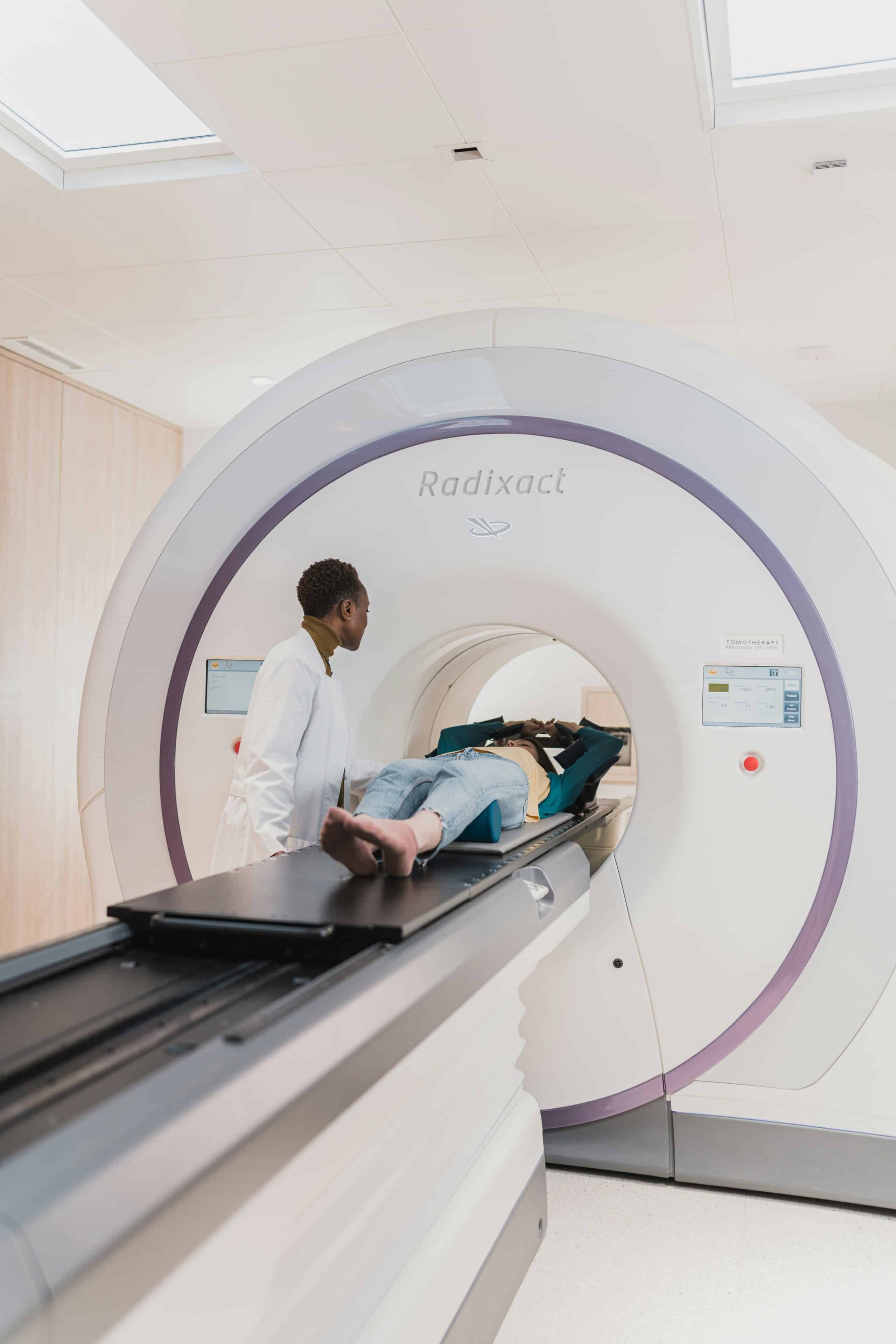
Persistent pelvic pain that does not respond to standard treatments can sometimes indicate underlying conditions such as endometriosis, a chronic and often underdiagnosed disorder that may take years to identify. While imaging plays an important role in evaluating pelvic pain, not all modalities are equally effective for detecting endometriosis. CT scans are not considered a primary diagnostic tool for endometriosis, but they can help identify complications or rule out other causes of symptoms as part of a broader clinical assessment.
In this guide, we explain what endometriosis is, how CT imaging works, and where it fits within the diagnostic process. We also compare CT scans with other imaging methods such as MRI and ultrasound, outline what patients can expect during the procedure, and review current clinical recommendations. By the end, you will have a clear understanding of the appropriate role of CT scans in endometriosis evaluation and when to seek care from a specialist experienced in managing this condition.
What Exactly Is Endometriosis and Why Is Pinpointing It Crucial?

Endometriosis is a chronic condition in which tissue similar to the lining of the uterus grows outside the uterus. These growths can occur on the ovaries, fallopian tubes, bowel, bladder, and other pelvic structures. Over time, this tissue can trigger inflammation, scarring, and adhesions, leading to persistent pelvic pain and other complications.
Accurate diagnosis is important because endometriosis presents differently in each individual and can overlap with other conditions. Identifying the condition early allows for more targeted treatment planning, which may include medical management, pain control strategies, or surgical intervention depending on disease severity and patient goals. Early diagnosis also supports better long-term outcomes, including fertility preservation and improved quality of life.
What Are the Common Signs and How Does Endometriosis Affect Daily Life?
Endometriosis symptoms can vary in intensity and may change over time. Common signs include:
- Chronic pelvic pain that may worsen during menstruation
- Severe menstrual cramps that interfere with daily activities
- Pain during or after intercourse
- Difficulty conceiving or infertility
These symptoms can significantly affect physical, emotional, and social well-being. Many individuals experience fatigue, reduced productivity, and increased stress or anxiety, particularly when symptoms are not clearly explained or effectively managed.
Why Is Endometriosis Difficult to Diagnose Early?
Early diagnosis of endometriosis can be challenging for several reasons:
- Symptom overlap with other conditions such as irritable bowel syndrome, pelvic inflammatory disease, or ovarian cysts
- Limitations of non-invasive imaging, which may not detect small or superficial lesions
- Variable disease presentation, with lesions occurring in multiple pelvic and sometimes extra-pelvic locations
Because of these factors, diagnosis often requires a combination of clinical evaluation, imaging, and in some cases, surgical assessment. This complexity contributes to delays in diagnosis for many patients.
How Does an Accurate Diagnosis Influence Treatment Outcomes?
A confirmed diagnosis allows clinicians to make more informed treatment decisions based on the extent and location of disease, as well as patient priorities.
- Personalized treatment planning: Options may include hormonal therapy, pain management, or surgical approaches such as excision of endometriosis lesions
- Fertility considerations: Early and appropriate intervention can help reduce the impact of scarring on reproductive organs
- Ongoing monitoring: Establishing a diagnosis provides a baseline for tracking symptoms and response to treatment over time
When care is guided by an accurate diagnosis and aligned with patient goals, individuals are more likely to experience meaningful symptom relief and avoid unnecessary or ineffective treatments.
What Is a CT Scan and How Is It Used in Evaluating Endometriosis?
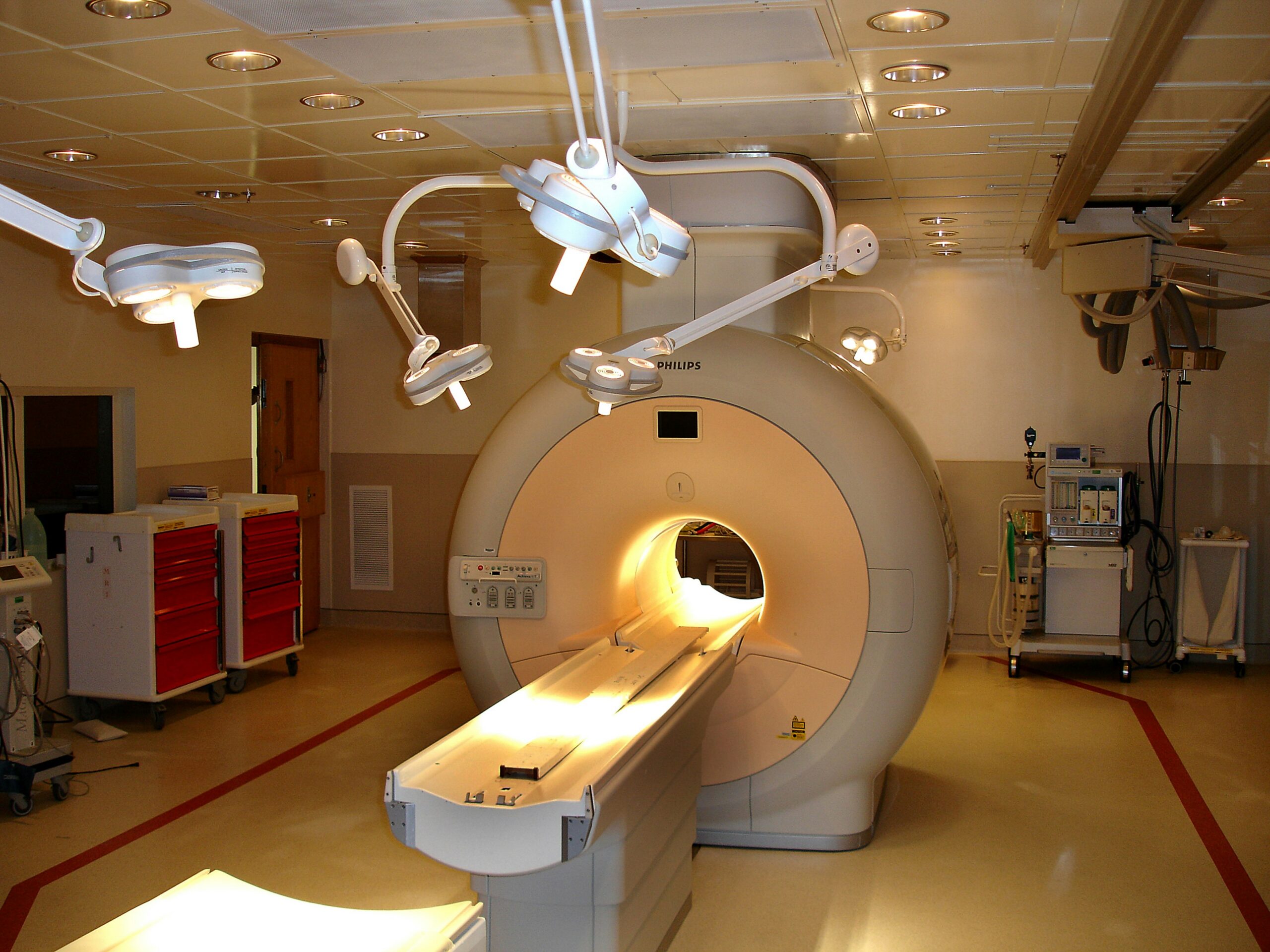
Computed Tomography (CT) is an imaging technique that uses X-rays and computer processing to create detailed cross-sectional images of the body. It is commonly used to evaluate structures within the abdomen and pelvis and can help identify a wide range of conditions that cause pelvic pain.
In the context of endometriosis, CT scans are not used as a primary diagnostic tool. However, they can play a supportive role by identifying complications or ruling out other conditions with similar symptoms. CT imaging is often used in emergency or complex clinical situations where a rapid and broad assessment is required.
How Does CT Imaging Work?
CT imaging involves a rotating X-ray source that captures multiple images from different angles around the body. These images are processed by a computer to create detailed cross-sectional views.
- X-ray beams pass through the body at multiple angles
- Detectors measure how different tissues absorb these beams
- Computer algorithms reconstruct the data into layered images
This process allows clinicians to evaluate organ structure and identify abnormalities such as fluid collections, masses, or signs of inflammation.
What Areas Are Evaluated on a CT Scan?
When assessing pelvic or abdominal symptoms, CT scans typically evaluate:
- Ovaries and adnexal regions: May reveal masses, though CT cannot reliably distinguish endometriomas from other cysts
- Bowel and bladder: Can show wall thickening, obstruction, or inflammatory changes
- Urinary tract: Useful for detecting hydronephrosis caused by ureteral compression
These findings can help determine whether additional imaging, such as MRI or ultrasound, or specialist referral is needed.
When Is a CT Scan Recommended for Suspected Endometriosis?
CT scans are usually considered in specific clinical scenarios rather than for routine diagnosis of endometriosis. These include:
- Acute or severe pelvic pain, where conditions such as appendicitis, diverticulitis, or abscess need to be ruled out
- Suspected urinary tract involvement, particularly to evaluate for ureteral obstruction and hydronephrosis
- Complex cases requiring broader evaluation, especially when bowel or other abdominal organs may be involved
By identifying complications or alternative diagnoses, CT scans can support clinical decision-making and help guide further evaluation or treatment planning within a multidisciplinary care setting.
Can a CT Scan Detect Endometriosis?
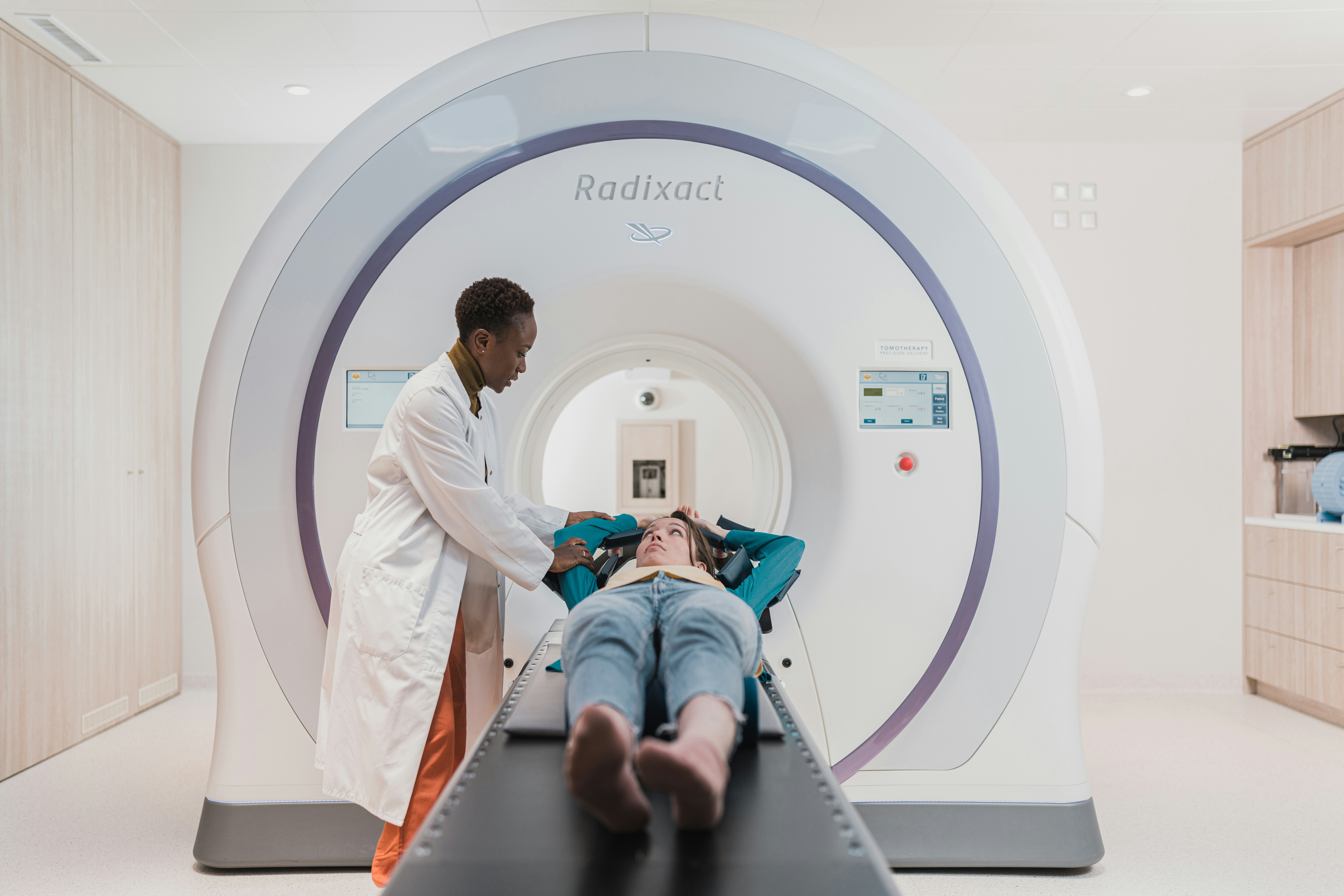
CT scans are not considered a reliable method for directly diagnosing endometriosis. The condition often involves small or superficial lesions that are difficult to visualize with CT due to its limited soft tissue contrast. As a result, CT imaging is not typically used to confirm the presence of endometriosis.
However, CT scans can still play a role in evaluation by identifying complications or excluding other conditions that may cause similar symptoms. This makes CT a supportive tool within a broader diagnostic approach rather than a standalone solution.
What Findings Related to Endometriosis May Appear on CT?
Although CT cannot reliably detect endometriosis itself, it may reveal indirect or associated findings, including:
- Adnexal masses: CT may identify ovarian cysts or masses, but it cannot reliably distinguish endometriomas from other types of cysts
- Bowel involvement: Findings such as wall thickening or surrounding inflammation may suggest possible involvement but are not specific to endometriosis
- Urinary tract changes: Hydronephrosis may be visible if endometriosis is compressing the ureters
These findings often prompt further evaluation with more sensitive imaging, such as MRI, or referral to a specialist.
How Effective Is CT for Detecting Deep Endometriosis?
CT has limited ability to detect deep infiltrating endometriosis. Lesions can blend with surrounding tissues, making them difficult to distinguish. Even with contrast enhancement, CT does not provide the level of soft tissue detail needed for accurate mapping of disease.
MRI is generally preferred for evaluating deep endometriosis because it offers superior soft tissue contrast and better visualization of lesion extent.
What Complications Are Best Seen on CT?
CT imaging is most useful when complications are suspected. It can help identify:
- Ureteral obstruction, including associated hydronephrosis
- Bowel obstruction, including the location and severity of blockage
- Inflammatory collections or abscesses within the pelvis or abdomen
In these situations, CT provides rapid and comprehensive assessment, which is particularly valuable in urgent or complex cases. We cover this further in Comprehensive Guide: Endometriosis Assessment Best Practices.
What Are the Limitations of CT in Endometriosis Diagnosis?
Several limitations restrict the role of CT in diagnosing endometriosis:
- Limited soft tissue contrast, reducing the ability to detect small or superficial lesions
- Non-specific findings, as many abnormalities seen on CT can be caused by conditions other than endometriosis
- Radiation exposure, which is an important consideration, particularly for individuals of reproductive age
Because of these limitations, CT is typically used alongside other diagnostic methods. MRI, ultrasound, and in some cases laparoscopy remain the primary tools for accurate diagnosis and disease assessment.
How Do CT Scans Compare to MRI, Ultrasound, and Laparoscopy in Diagnosing Endometriosis?
Different diagnostic methods provide distinct types of information when evaluating endometriosis. Understanding how each modality performs helps guide appropriate clinical decision-making based on symptoms, suspected disease extent, and patient priorities.
Comparison of Imaging Modalities
Each method serves a different purpose. CT is primarily used to identify complications or exclude other causes of pelvic pain. MRI is preferred for detailed mapping of disease. Ultrasound is often used as a first-line imaging tool, particularly for evaluating ovarian involvement. Laparoscopy remains the definitive method when confirmation is required. For more detail, see our article Why Endometriosis Pain Often Goes Undiagnosed for Years.
Why Is MRI More Effective Than CT for Endometriosis?
MRI provides superior soft tissue contrast compared to CT, allowing for more accurate visualization of deep infiltrating endometriosis, adhesions, and involvement of surrounding structures. It can evaluate disease in multiple planes and identify features such as blood-containing lesions with greater clarity.
Because MRI does not use ionizing radiation and offers higher diagnostic accuracy for mapping disease extent, it is generally preferred when endometriosis is strongly suspected. You may also find What Influences the Accuracy of Endometriosis Diagnoses helpful.
What Advantages Does Ultrasound Offer?
Transvaginal ultrasound is widely used as an initial imaging tool because it is accessible, cost-effective, and does not involve radiation.
Key advantages include:
- Real-time imaging that allows dynamic assessment
- Reliable detection of ovarian endometriomas, often with characteristic internal appearance
- Evaluation of pelvic organs, particularly the ovaries and anterior compartment
However, its ability to detect deep infiltrating endometriosis depends on operator expertise and lesion location.
Why Is Laparoscopy Still Considered the Gold Standard?
Laparoscopy remains the reference standard for diagnosing endometriosis because it allows direct visualization of lesions and confirmation through biopsy. It also enables treatment during the same procedure, such as excision of endometriosis tissue.
Although imaging techniques have improved, they cannot consistently detect all forms of the disease, especially superficial lesions. For this reason, laparoscopy is often used when symptoms persist despite inconclusive imaging or when surgical treatment is being considered.
Key Strengths and Limitations of Each Approach
- CT Scan: Useful for detecting complications and excluding other diagnoses, but limited in identifying endometriosis lesions
- MRI: Provides detailed mapping of disease and is preferred for evaluating deep involvement
- Ultrasound: Effective first-line tool for ovarian disease, but less consistent for deeper lesions
- Laparoscopy: Offers definitive diagnosis and treatment, but involves surgical risks
The choice of diagnostic approach depends on clinical presentation, available resources, and whether the goal is initial evaluation, detailed mapping, or definitive confirmation.
What Should Patients Expect During a CT Scan for Pelvic Pain?
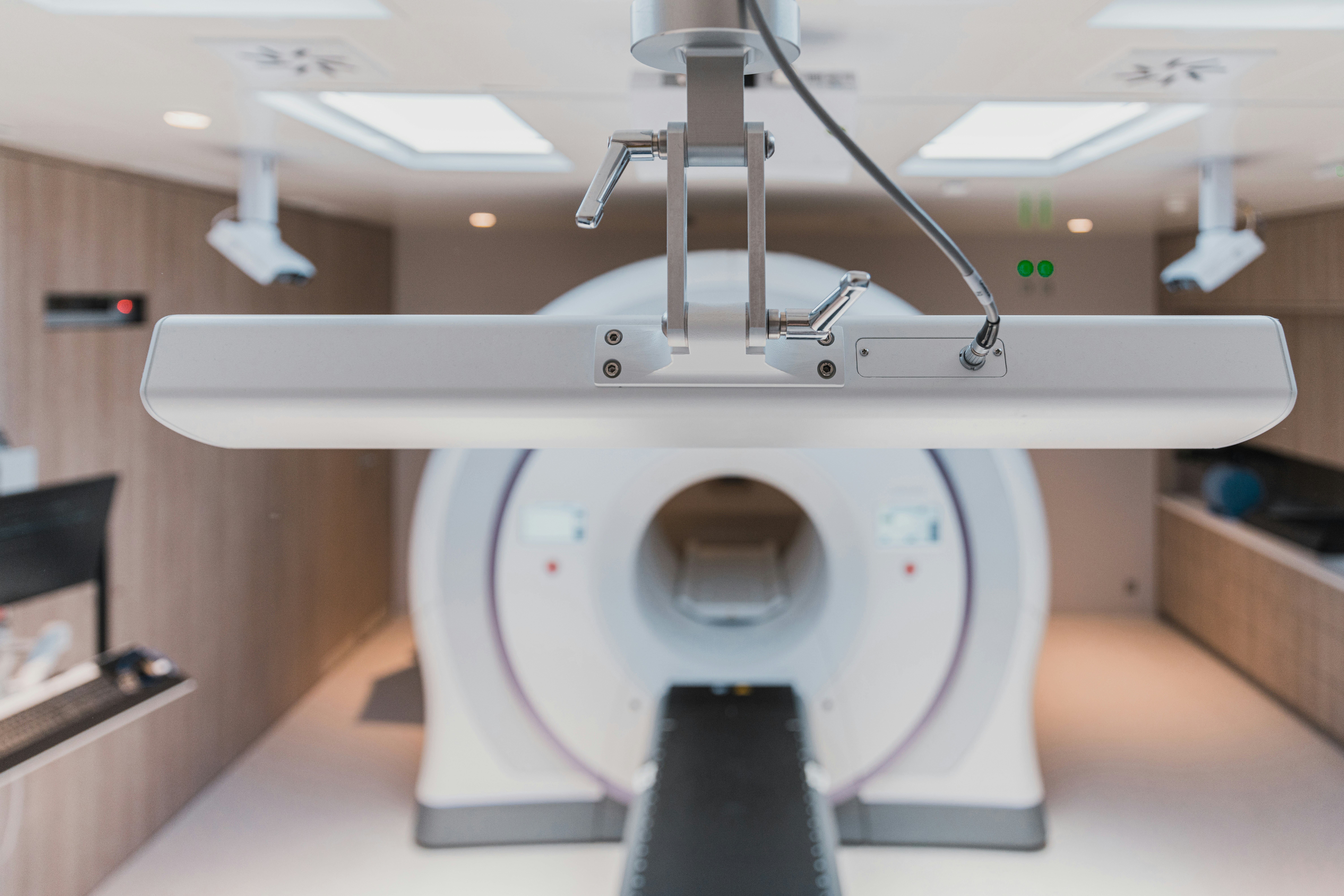
A CT scan of the abdomen and pelvis is a quick and commonly performed imaging test used to evaluate unexplained pelvic pain and identify possible complications. While it is not used to diagnose endometriosis directly, it can help assess other potential causes of symptoms and guide further evaluation.
Understanding the procedure can help reduce uncertainty and make the experience more comfortable.
How Is a CT Scan Performed?
During the scan, the patient lies on a motorized table that moves through a circular scanner. The procedure is painless and typically completed within a few minutes.
- The patient remains still while images are taken
- The scanner rotates around the body to capture images from multiple angles
- In some cases, an intravenous contrast agent is used to improve visualization of blood vessels and areas of inflammation
Most scans are completed within 5 to 10 minutes, although preparation time may vary slightly depending on whether contrast is required.
Are There Any Risks or Contraindications?
CT scans are generally safe, but there are some considerations:
- Radiation exposure: CT uses ionizing radiation, so use is carefully considered, especially in younger patients or those requiring repeated imaging
- Contrast-related risks: Some patients may experience mild allergic reactions, and kidney function may need to be assessed before contrast is given
- Pregnancy: CT is usually avoided during pregnancy unless absolutely necessary; alternative imaging methods are preferred
Healthcare providers weigh these factors when deciding whether a CT scan is appropriate.
How Should Patients Prepare?
Preparation depends on whether contrast is used, but general recommendations include:
- Avoid eating or drinking for several hours before the scan if contrast is planned
- Inform the care team about allergies, medications, pregnancy status, or kidney conditions
- Wear comfortable clothing and remove metal objects such as jewelry
Following these instructions helps ensure clear imaging and reduces the likelihood of needing repeat scans.
What Happens After the Scan?
After the procedure, patients can usually return to normal activities immediately. If contrast was used, hydration may be recommended to help clear it from the body. A radiologist reviews the images and sends a report to the referring clinician, who will discuss the results and next steps.
What Are the Latest Research Findings and Guidelines on Using CT Scans for Endometriosis Diagnosis?
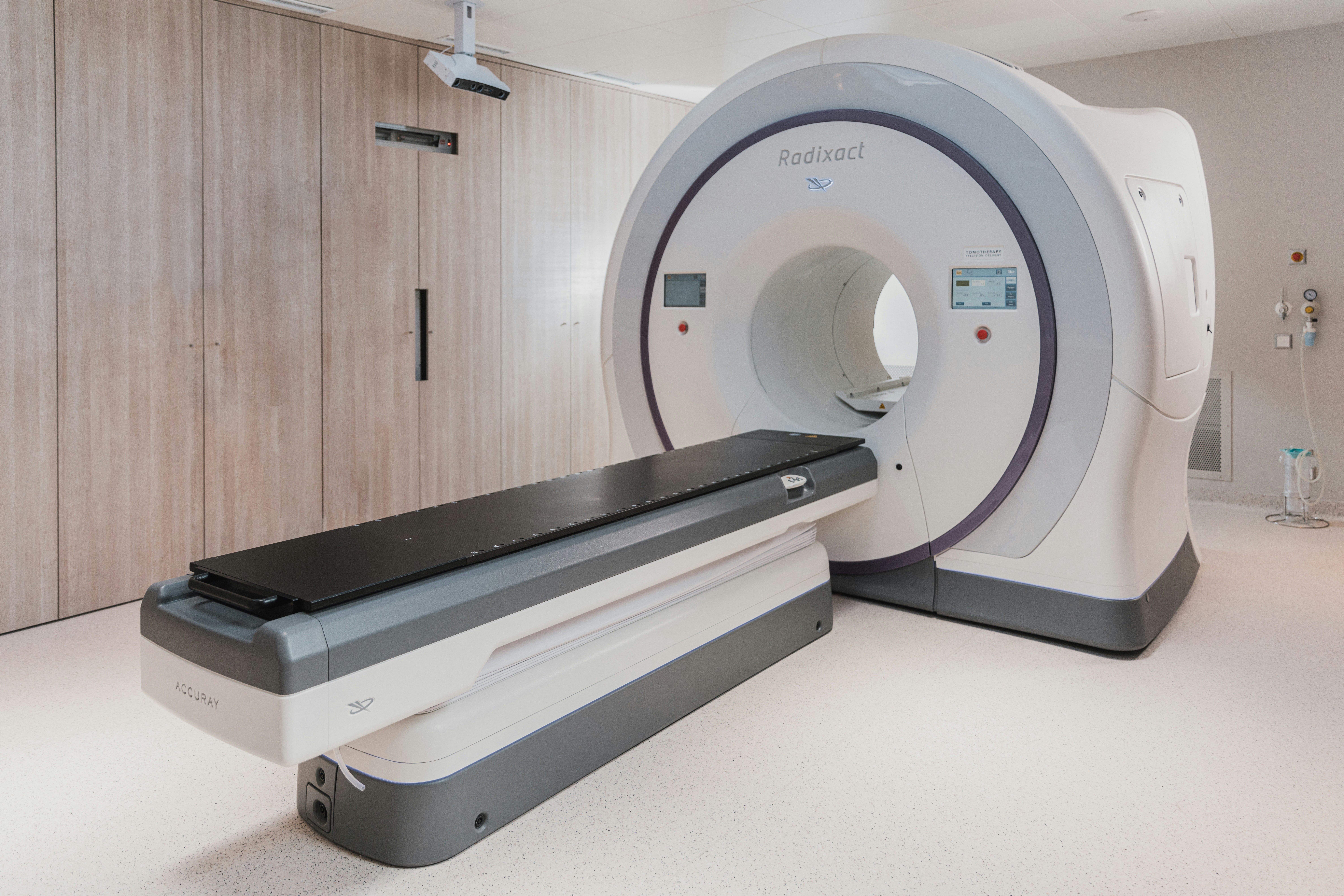
Current research and clinical guidelines define a limited but specific role for CT scans in the evaluation of suspected endometriosis. While CT is widely used in general abdominal imaging, it is not recommended as a primary diagnostic tool for endometriosis due to its inability to reliably detect small or superficial lesions.
Instead, CT is used selectively to evaluate complications or to rule out other causes of pelvic pain. MRI, ultrasound, and laparoscopy remain the preferred methods for diagnosing and assessing the extent of disease.
What Does Research Indicate About CT Scan Accuracy?
Evidence consistently shows that CT has low sensitivity for detecting endometriosis lesions, particularly superficial and small implants. Even in cases of deeper disease, CT may not clearly distinguish abnormal tissue from surrounding structures.
However, CT can be useful in identifying complications associated with advanced disease, such as:
- Urinary tract obstruction and hydronephrosis
- Bowel involvement or obstruction
- Inflammatory changes within the abdomen or pelvis
Because of these limitations, CT is typically used as part of a broader diagnostic strategy rather than as a standalone tool.
How Do Clinical Guidelines Recommend Using CT?
Guidelines from major gynecological and radiological organizations generally recommend:
- Using ultrasound or MRI as first-line imaging when endometriosis is suspected
- Reserving CT for acute or unclear cases, particularly when alternative diagnoses need to be excluded
- Using CT selectively when evaluating complications involving the bowel or urinary tract
- Avoiding routine CT use for endometriosis diagnosis due to limited diagnostic value and radiation exposure
This approach ensures that imaging is both clinically appropriate and aligned with patient safety considerations.
What Are Emerging Developments in Imaging?
Advances in imaging technology are being explored to improve detection and characterization of endometriosis, although many are still in early stages of clinical use.
- Advanced CT techniques, such as dual-energy CT, are being studied for improved tissue differentiation
- Artificial intelligence applications are under development to assist in identifying subtle imaging patterns
- Multimodal imaging approaches continue to evolve, combining different techniques to improve diagnostic accuracy
While these developments are promising, they are not yet standard practice. MRI and ultrasound remain the most reliable non-invasive imaging tools, with laparoscopy used for definitive diagnosis when needed.
Frequently Asked Questions
What lifestyle changes can help manage endometriosis symptoms?
Managing endometriosis symptoms often involves lifestyle modifications. Regular exercise can help reduce pain and improve overall well-being. A balanced diet rich in anti-inflammatory foods, such as fruits, vegetables, and omega-3 fatty acids, may alleviate symptoms. Additionally, stress management techniques like yoga, meditation, or counseling can be beneficial. It’s essential to consult with a healthcare provider for personalized recommendations that align with your specific condition and needs. You may also find Understanding Endometriosis Pain Assessment Techniques helpful.
Are there any alternative therapies for endometriosis?
Many individuals explore alternative therapies to complement traditional treatments for endometriosis. Acupuncture, for instance, has shown promise in reducing pelvic pain and improving quality of life. Herbal supplements, such as ginger and turmeric, may also provide anti-inflammatory benefits. However, it’s crucial to discuss any alternative therapies with a healthcare professional to ensure they are safe and effective in conjunction with your current treatment plan.
How does endometriosis affect mental health?
Endometriosis can significantly impact mental health due to chronic pain, fertility challenges, and the emotional toll of living with a chronic condition. Many individuals experience anxiety, depression, and feelings of isolation. It’s essential to address these mental health concerns through counseling, support groups, or therapy. Open communication with healthcare providers about mental health can lead to a more comprehensive treatment approach that includes both physical and emotional support.
What role does diet play in managing endometriosis?
Diet can play a crucial role in managing endometriosis symptoms. Some studies suggest that a diet low in red meat and high in fruits, vegetables, and whole grains may help reduce inflammation and pain. Foods rich in omega-3 fatty acids, such as fish and flaxseeds, are also beneficial. Keeping a food diary to track symptoms can help identify potential dietary triggers. Consulting a nutritionist familiar with endometriosis can provide tailored dietary advice.
What are the long-term effects of untreated endometriosis?
Untreated endometriosis can lead to several long-term complications, including chronic pain, infertility, and the formation of adhesions that can affect organ function. Over time, the condition may worsen, leading to more severe symptoms and complications such as bowel or bladder issues. Early diagnosis and treatment are crucial to managing the disease effectively and preventing these long-term effects. Regular follow-ups with a healthcare provider are essential for monitoring the condition.
How can I find support for living with endometriosis?
Finding support for living with endometriosis is vital for emotional and psychological well-being. Many organizations and online communities offer resources, forums, and support groups where individuals can share experiences and coping strategies. Local support groups may also be available through hospitals or clinics. Connecting with others who understand the challenges of endometriosis can provide comfort and valuable insights into managing the condition.
Conclusion
Understanding the role of CT scans in diagnosing endometriosis is essential for effective patient care, as they help identify complications and rule out other conditions. While CT imaging has its limitations, it plays a vital part in a comprehensive diagnostic strategy alongside MRI and laparoscopy. For those seeking expert guidance and personalized treatment plans, the Endo Excellence Center is here to support you. Explore our resources and connect with our team to take the next step towards managing your health. When you are ready to talk through your options, schedule an appointment with our team.