
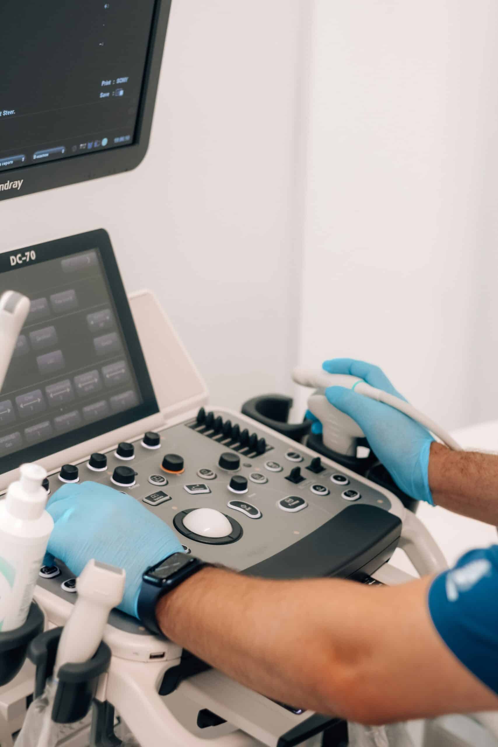
Endometriosis affects one in ten women of reproductive age, yet a diagnosis often takes nearly a decade. Advanced, non-invasive imaging techniques can revolutionize how we plan treatment and improve outcomes. This guide explores the science behind transvaginal ultrasound and pelvic MRI, how they work together, the vital role of expert interpretation, exciting AI advancements, and a clear path for patients from initial symptoms to surgical readiness. Understanding these sophisticated methods will empower you to navigate your diagnostic journey with confidence and partner with specialized centers for personalized care.
What Is Endometriosis and Why Is Precise Imaging So Important?
Endometriosis is a chronic condition where tissue similar to the uterine lining grows outside the uterus, leading to pelvic pain, painful periods, and infertility. Accurate imaging allows us to visualize these lesions, map deep infiltrating endometriosis, and tailor treatment without immediately resorting to surgery. For instance, identifying an ovarian endometrioma on ultrasound helps us plan the most effective medical or surgical approach before symptoms worsen.
What Are the Common Forms and Signs of Endometriosis?
Endometriosis typically appears in three main forms: superficial peritoneal implants, endometriomas (cysts on the ovaries), and deep infiltrating endometriosis (DIE). Each can cause unique symptoms and complications.
- Experience pelvic pain that becomes more intense during your period and lingers afterward.
- Notice pain during intercourse (dyspareunia) if lesions have infiltrated sensitive areas.
- Observe digestive or urinary issues if lesions affect the bowel or bladder.
These symptoms often overlap, making imaging essential to pinpoint the exact type and location of lesions, which directly guides the best diagnostic and treatment choices.
Why Does Endometriosis Diagnosis Often Take So Long?
Diagnosis frequently takes seven to ten years because symptoms can mimic other conditions, and physical exams may not detect hidden lesions.
- You might need to see several specialists before imaging is ordered.
- Symptom severity can vary greatly, leading some to believe their pain is normal.
- Pelvic exams can only identify superficial nodules and masses.
To shorten this delay, we need earlier imaging referrals and standardized ultrasound or MRI protocols to uncover hidden disease.
How Does Imaging Help Detect Endometriosis Better Than Physical Exams?
Imaging allows us to see lesions that are impossible to feel during a manual exam. It helps identify endometriomas and deep infiltrating disease, leading to better treatment planning and reducing the need for invasive laparoscopy.
- Provides a real-time view of ovarian cysts, adhesions, and nodules.
- Maps the depth of lesions and which organs are involved for accurate staging.
- Offers detailed anatomical guidance for surgical planning.
By revealing what’s hidden, imaging significantly improves diagnostic accuracy and your confidence in receiving personalized care.
How Does Transvaginal Ultrasound Help Detect Endometriosis?

Transvaginal ultrasound uses a specialized transducer placed inside the vagina to send sound waves into the pelvic organs. These waves bounce back, creating detailed images that reveal lesions, endometriomas, and adhesions. This non-invasive method speeds up diagnosis and helps us create targeted treatment plans.
What Is Transvaginal Ultrasound and How Is It Used for Endometriosis?
Transvaginal ultrasound involves a slim probe that emits sound pulses. As these pulses echo off tissues, they form live images of your uterus, ovaries, and pelvic ligaments. By examining how these echoes appear and the shape of lesions, sonographers can identify endometriomas—often seen as uniform, darker cysts with a “ground-glass” look—and detect deep nodules affecting nearby structures.
How Well Does Ultrasound Detect Endometriomas and Deep Infiltrating Endometriosis?
Ultrasound is highly accurate for endometriomas and can detect DIE in 70–80% of cases when performed by experienced professionals.
- It can identify ovarian cysts larger than 1 cm and clearly visualize their walls.
- It can detect infiltration of the rectosigmoid and uterosacral ligaments in many advanced cases.
- It can help distinguish endometriomas from other types of cysts using Doppler flow imaging.
Skilled technique and advanced equipment lead to reliable detection, enabling earlier intervention and better patient outcomes.
What Do I Need to Do to Prepare for an Endometriosis Ultrasound?
Proper preparation ensures the clearest possible images and the most accurate diagnosis.
- Empty your bladder before the exam so the probe can be positioned optimally.
- Follow any bowel preparation instructions, which might include a light meal or prescribed laxatives.
- Schedule your exam during the early part of your menstrual cycle (days 5–10) for the best view of your ovaries.
Following these steps helps ensure precise mapping of lesions and reduces the need for repeat scans, making your diagnostic process smoother.
What Are the Pros and Cons of Using Ultrasound for Endometriosis Diagnosis?
The table below highlights the key features, benefits, and considerations of ultrasound to help you understand its role in diagnosis.
This comparison shows that ultrasound is a valuable first-line tool, while MRI offers additional benefits for comprehensive staging.
What Role Does Pelvic MRI Play in Diagnosing and Planning Surgery for Endometriosis?

Pelvic MRI uses magnetic fields and radio waves to create detailed cross-sectional images of your pelvic organs. It offers superior contrast, allowing us to precisely map deep infiltrating endometriosis, adhesions, and lesions outside the pelvis, which is crucial for effective surgical planning.
How Does Pelvic MRI Detect Deep Infiltrating Endometriosis and Other Lesions?
Pelvic MRI identifies DIE by highlighting areas of blood (hyperintense on T1-weighted images) and scar tissue (hypointense on T2-weighted images). It can reveal nodules in the uterosacral ligaments, rectovaginal septum, and bowel wall, guiding surgeons to the exact locations and showing the depth of infiltration needed for precise excision.
When Is MRI a Better Choice Than Ultrasound for Endometriosis?
MRI is recommended when ultrasound can’t fully show the extent of the disease or when endometriosis might be affecting areas outside the pelvis.
- To assess potential involvement of the bowel, bladder, or ureters, which may be beyond the scope of ultrasound.
- To clarify complex anatomy in cases of recurrent or widespread endometriosis.
- For detailed pre-operative staging to guide multidisciplinary surgical teams.
Choosing MRI in these situations leads to more accurate lesion mapping and fewer surprises during surgery.
How Should Patients Prepare for an Endometriosis MRI Scan?
Proper preparation helps ensure the best possible image quality and your comfort during the scan.
- Avoid eating for four to six hours beforehand to minimize bowel movement artifacts.
- You may receive medication to slow bowel movements, reducing motion blur.
- If bowel wall involvement is a key concern, a gel may be instilled into the vagina or rectum.
Following these steps enhances lesion visibility and supports accurate interpretation.
What Are the Advantages and Limitations of MRI for Endometriosis Imaging?
This overview highlights MRI’s strength as a detailed diagnostic tool for comprehensive disease assessment and surgical strategy.
How Do Ultrasound and MRI Compare for Endometriosis Diagnostic Imaging?
Both ultrasound and MRI play crucial, complementary roles in diagnosing endometriosis, leading to more accurate assessments and better patient care.
What Are the Complementary Roles of Ultrasound and MRI in Endometriosis Diagnosis?
Often, ultrasound is used first to look for endometriomas and superficial disease. MRI is then used for detailed mapping of unclear or deep lesions. Together, they offer a balance of accessibility, cost-effectiveness, and thorough disease staging.
Which Imaging Technique Is Best for Different Types of Endometriosis?
This guide helps tailor imaging choices based on the suspected type and location of endometriosis.
How Does Imaging Accuracy Impact Treatment and Surgical Planning?
More accurate imaging leads to more precise treatment decisions, reducing the need for exploratory surgery and ensuring that all lesions are addressed during excision. Detailed mapping of nodules and adhesions can shorten surgery, lower complication rates, and decrease the risk of recurrence by enabling coordinated multidisciplinary care.
What Are the Key Differences in Patient Experience Between Ultrasound and MRI?
Patients generally find ultrasound to be quicker and less confining. MRI provides more detailed information but involves longer scan times and can sometimes be uncomfortable. Understanding these differences helps us prepare patients better and ensure they are comfortable throughout the diagnostic process.
Why Is Expert Interpretation Essential for Endometriosis Imaging?

Having an expert interpret your imaging is crucial to ensure that even subtle endometriotic lesions, especially those deep within tissues, are identified. This expert analysis directly informs the best surgical and treatment strategies.
What Role Do Specialized Sonographers and Radiologists Play in Accurate Diagnosis?
Specialized sonographers use advanced techniques, like specific bowel preparation and detailed DIE scanning protocols. Expert radiologists then combine these imaging findings with surgical anatomy knowledge. This collaboration helps identify lesion characteristics that might be missed by general practitioners.
How Does Expert Imaging Interpretation Improve Surgical Outcomes?
Accurate interpretation minimizes unexpected findings during surgery, reduces operating time, and helps ensure complete removal of lesions. By anticipating the location of adhesions and how they might affect surrounding structures, surgeons can plan less invasive procedures and better preserve fertility.
How Does EndoExcellenceCenter Ensure High-Quality Imaging and Interpretation?
At Endo Excellence Center, our team is committed to excellence through:
- Rigorous training for our sonographers in standardized, consensus-based scanning protocols.
- Regular multidisciplinary meetings involving gynecologic surgeons and radiologists to review complex cases.
- Utilizing state-of-the-art high-resolution ultrasound machines and 3T MRI scanners for exceptional image clarity.
These measures provide diagnostic confidence and support the development of personalized surgical plans.
What Emerging Technologies Are Shaping the Future of Endometriosis Imaging?

Exciting advancements in artificial intelligence, new imaging agents, and improved scanning sequences are poised to shorten diagnostic timelines and enhance lesion detection beyond current capabilities.
How Is Artificial Intelligence Enhancing Endometriosis Diagnostic Imaging?
AI algorithms can analyze ultrasound and MRI data to spot subtle lesion patterns, measure disease extent, and predict involvement of specific bowel segments. By automating the identification and classification of lesions, AI can speed up reporting and ensure more consistent staging.
What New Imaging Agents and Techniques Are Being Developed?
Researchers are exploring specialized MRI agents designed to highlight endometriotic tissue and fluorescent dyes for use during surgery to better visualize lesions. Additionally, techniques like elastography are being developed to assess tissue stiffness, helping to differentiate fibrotic implants from healthy tissue.
How Will These Advances Reduce Diagnostic Delays and Improve Patient Care?
By improving our ability to detect lesions, especially early or superficial ones, these emerging technologies will allow for earlier treatment, more personalized medical therapies, and fewer invasive procedures. As imaging becomes even more precise, patient outcomes and quality of life will significantly improve.
How Can Patients Navigate the Endometriosis Diagnostic Imaging Journey?
Understanding when to get imaging, how to interpret the results, and what to expect next empowers you to take an active role in your care.
When Should You Seek Imaging for Endometriosis Symptoms?
Consider imaging if you are experiencing:
- Persistent pelvic pain that doesn’t improve with initial treatments.
- Digestive or urinary issues that occur around your menstrual cycle.
- Difficulty conceiving after six months of trying without contraception.
Seeking imaging early can lead to a faster diagnosis and prevent symptoms from worsening.
How to Understand and Interpret Your Endometriosis Imaging Report?
Pay attention to key details in your report, such as lesion location, size, depth, and which organs are affected. Discuss the surgical mapping recommendations with your doctor. Don’t hesitate to ask for explanations of any technical terms and how the findings will shape your treatment options.
What Are the Next Steps After Imaging: Treatment and Surgical Planning?
Following your imaging, your care team will:
- Suggest medical treatments (like hormone therapy or pain management) if lesions are minor.
- Plan minimally invasive surgery (laparoscopy or excision) for deep or symptomatic disease.
- Arrange consultations with multiple specialists for complex cases involving the bowel or bladder.
This structured approach ensures a smooth transition from diagnosis to effective treatment.
Where to Find Support and Resources During Your Diagnostic Process?
Look for support through:
- Local endometriosis support groups and online communities.
- Educational webinars offered by professional organizations (like ESHRE or ISUOG).
- Patient navigators at specialized centers who can help coordinate appointments and offer guidance.
Accessing these resources can reduce anxiety and help you make informed decisions at every stage.
Don’t let pelvic pain and fertility concerns remain unanswered. Advanced imaging provides a clear path toward personalized care. Transvaginal ultrasound and MRI work together to create a comprehensive diagnostic picture, while expert interpretation and emerging AI tools drive precision and innovation. At Endo Excellence Center, our specialized team and cutting-edge protocols ensure you receive timely, accurate insights that guide optimal treatment planning. Embrace your diagnostic journey with confidence, knowing that expert-guided imaging leads the way to relief and a better quality of life.
Frequently Asked Questions
How long does it typically take to get an endometriosis diagnosis?
Diagnosis often takes seven to ten years because symptoms can mimic other conditions and physical exams cannot detect hidden lesions. Advanced imaging, such as transvaginal ultrasound and pelvic MRI, can shorten this delay significantly by revealing lesions that a manual exam would miss.
What is the difference between transvaginal ultrasound and pelvic MRI for endometriosis?
Transvaginal ultrasound is typically used first to identify endometriomas and superficial disease, offering real-time, cost-effective imaging. Pelvic MRI provides superior soft-tissue contrast and is better suited for mapping deep infiltrating endometriosis, extra-pelvic lesions, and complex anatomy ahead of surgery.
Do I need to prepare for an endometriosis ultrasound or MRI?
Yes. For ultrasound, patients are generally asked to empty their bladder beforehand and schedule the exam during days 5 to 10 of their cycle. For MRI, patients typically avoid eating for four to six hours prior and may receive medication or a gel instillation to reduce motion artifacts and improve lesion visibility.
Can imaging alone confirm an endometriosis diagnosis?
Imaging is a powerful diagnostic tool that can identify endometriomas and map deep infiltrating disease with high accuracy, but interpretation by an experienced sonographer or radiologist is essential. Expert interpretation ensures subtle lesions are not missed and directly informs the most effective treatment and surgical strategy.
How does Endo Excellence Center ensure accurate imaging and interpretation?
Endo Excellence Center relies on rigorously trained sonographers following standardized, consensus-based scanning protocols, regular multidisciplinary case reviews between gynecologic surgeons and radiologists, and state-of-the-art high-resolution ultrasound and 3T MRI equipment to deliver reliable, personalized diagnostic insight.
Conclusion
Endometriosis is a complex condition, but precise imaging transforms the diagnostic journey from years of uncertainty into a clear, actionable path forward. Transvaginal ultrasound and pelvic MRI each bring distinct strengths, and when combined with expert interpretation, they give physicians the detailed picture needed to plan effective medical or surgical care. As AI-driven analysis and new imaging techniques continue to advance the field, patients can look forward to faster diagnoses and even more personalized treatment. At Endo Excellence Center, our commitment to rigorous protocols and multidisciplinary expertise ensures every patient receives the accurate, compassionate care needed to move confidently from symptoms to relief.