
A pelvic MRI can detect and map many forms of deep endometriosis, particularly when lesions affect the bowel, bladder, ureters, uterosacral ligaments, rectovaginal region, ovaries, or multiple pelvic compartments. It can also show fibrosis, organ tethering, endometriomas, adenomyosis, and other structural findings that may influence treatment planning.
However, MRI cannot identify every endometriosis lesion. Superficial implants and some small or subtle areas of deep disease may not appear clearly on the scan. A normal MRI therefore does not prove that endometriosis is absent, nor does an abnormal scan automatically mean that surgery is required.
The usefulness of MRI depends on more than the scanner itself. The examination must address the correct clinical question, use an appropriate pelvic endometriosis protocol, and be interpreted by a radiologist familiar with the disease. NICE recommends specialist transvaginal ultrasound or pelvic MRI to diagnose and assess the extent of suspected deep endometriosis. It also states that these examinations should be planned and interpreted by professionals with specialist gynecologic imaging expertise.
The term deep infiltrating endometriosis, often abbreviated as DIE, is still widely used. Current guidelines and radiology literature also commonly use the shorter term deep endometriosis. Both describe endometriosis that extends beneath the surface of affected tissue and may involve pelvic organs or supportive structures.
When Does a Pelvic MRI Add Useful Information?

MRI is not automatically required for every patient with painful periods, chronic pelvic pain, or suspected endometriosis. Current diagnostic pathways generally use clinical evaluation and transvaginal ultrasound as important early steps. MRI is more often used when the care team needs additional anatomical detail, particularly when deep or complex disease is suspected.
NICE recommends offering transvaginal ultrasound to patients with suspected endometriosis, even when the pelvic examination is normal. Specialist ultrasound or MRI may then be considered to diagnose deep endometriosis and assess its extent.
When Deep or Multiorgan Disease Is Suspected
MRI may be useful when symptoms, examination findings, or previous imaging raise concern for disease involving structures such as the:
-
Rectum or sigmoid colon
-
Rectovaginal space
-
Uterosacral ligaments
-
Bladder
-
Ureters
-
Ovaries
-
Pelvic sidewalls
-
Posterior cul-de-sac
-
Multiple pelvic compartments
Symptoms cannot identify the exact location of a lesion. However, cyclical pain with bowel movements, urinary symptoms that change with the menstrual cycle, deep pain during intercourse, an ovarian endometrioma, or findings suggesting reduced organ mobility may justify more detailed imaging.
MRI offers a broad view of the pelvis and can evaluate fibrosis, tethering, and distortion across multiple anatomical compartments. Specialist transvaginal ultrasound may provide complementary dynamic information, including organ mobility and site-specific tenderness.
When Ultrasound Results Are Inconclusive or Incomplete
MRI may be considered when:
-
Routine ultrasound does not explain significant symptoms
-
An ovarian or adnexal mass requires further characterization
-
Ultrasound suggests deep disease but does not show its full extent
-
The suspected area is difficult to evaluate through ultrasound
-
Pain or anatomy limits the ultrasound examination
-
Previous surgery has altered the normal pelvic anatomy
-
Multifocal disease is suspected
MRI should not be described as automatically superior to ultrasound. When specialist transvaginal ultrasound is performed by an experienced clinician, it can identify ovarian endometriomas and many forms of deep endometriosis, including disease involving the bowel, bladder, or ureters. The most useful test depends on the suspected location, available expertise, patient tolerance, and information needed for treatment planning.
When Surgery Is Being Considered
When surgery is under consideration, MRI can help estimate the likely location and extent of visible disease before the procedure. The scan may identify findings that suggest possible involvement of the bowel, bladder, ureters, ovaries, or several pelvic compartments.
This information can help the surgeon determine whether additional planning or collaboration may be needed. For example, suspected bowel or urinary tract involvement may influence whether colorectal or urologic expertise should be available.
MRI supports preparation, but it does not create a complete surgical map. Some lesions found during surgery may not have been visible on imaging, while some abnormalities seen on MRI may appear different when directly evaluated.
At the Endometriosis Center of Excellence, Dr. Rachael Haverland evaluates symptoms, imaging, previous treatment, bowel and bladder concerns, fertility goals, and surgical history together when determining whether minimally invasive excision surgery or another approach may be appropriate. The center also coordinates with other surgical disciplines when complex organ involvement is anticipated.
When MRI May Not Be the Immediate Next Step
MRI may not add meaningful information when:
-
Clinical assessment and ultrasound have not yet been completed
-
A recent specialist ultrasound has already answered the relevant anatomical questions
-
The MRI result would not change current management
-
The primary concern is superficial disease that may not be visible on MRI
-
The patient has a new acute symptom requiring another form of urgent evaluation
The goal is not to obtain the greatest possible number of tests. Imaging is most useful when it addresses a specific unanswered question and has the potential to change referral, monitoring, medical treatment, or surgical planning.
A Dedicated Endometriosis MRI Is Different From a Routine Pelvic MRI

Not every pelvic MRI is designed to evaluate deep endometriosis. A routine study may provide useful information about the uterus and ovaries but may not use the anatomical coverage, image orientation, or systematic reporting approach needed to assess all relevant pelvic compartments.
Radiology consensus guidance recommends a compartment-based approach to interpreting endometriosis imaging. It also emphasizes the importance of evaluating hemorrhagic lesions, fibrosis, anatomical distortion, and organ tethering rather than looking only for a visible mass.
The Referral Should State the Clinical Question
The imaging order should explain why endometriosis is suspected and identify the structures or symptoms that require particular attention.
Useful clinical information may include:
-
Cyclical bowel symptoms
-
Urinary pain or urgency associated with menstruation
-
Deep pain during intercourse
-
A known or suspected endometrioma
-
Previous endometriosis surgery
-
Possible bowel, bladder, or ureteral disease
-
Prior inconclusive ultrasound
-
Planned surgical evaluation
A clear referral helps the radiology team tailor the examination and interpret findings within the patient’s clinical context.
Patients should also bring or make available relevant previous ultrasound images, MRI studies, operative reports, and pathology records when requested. Comparing studies can help clarify whether a finding is new, stable, or related to prior treatment.
Endometriosis Imaging Requires Specific MRI Sequences
A dedicated MRI examination typically uses several image types and orientations to assess the pelvis. High-resolution T2-weighted images help show pelvic anatomy, fibrosis, tissue thickening, and distortion. T1-weighted images with fat suppression help identify blood-containing lesions, including many ovarian endometriomas.
MRI interpretation should not depend on a single finding. Deep endometriosis may appear through a combination of nodular tissue, low-signal fibrosis, organ retraction, tethering, and loss of normal tissue planes. The presence of visible blood products is not required for deep disease to be present.
Patients do not need to understand every technical sequence. The more practical question is whether the study was performed specifically to evaluate suspected endometriosis and whether all clinically relevant pelvic compartments were assessed.
Preparation Requirements Vary by Imaging Center
Instructions may differ among imaging centers. Depending on institutional practice and the clinical question, preparation may include:
-
Temporary fasting
-
Partial bladder filling
-
Medication intended to reduce bowel movement during the scan
-
Bowel preparation
-
Vaginal or rectal gel
-
Intravenous contrast in selected cases
None of these measures is universally required for every pelvic MRI. Patients should follow the instructions provided by the imaging center rather than using preparation guidance from another institution or an online source.
Anyone who may be pregnant, has kidney disease, has experienced a previous contrast reaction, or has an implanted medical device should notify the imaging team before the examination.
Contrast Is Not Automatically Necessary for Every Examination
Many characteristic findings of endometriosis can be evaluated through noncontrast T1- and T2-weighted sequences. Contrast may be used when the radiologist needs to assess an indeterminate ovarian mass, distinguish endometriosis from another condition, or answer an additional diagnostic question.
The decision to use contrast depends on the imaging protocol and the individual clinical situation. Patients should not assume that an MRI without contrast is incomplete or that every endometriosis scan requires contrast.
Radiologist Experience Influences the Value of the Scan
A technically adequate MRI can still be less informative when it is not interpreted by someone experienced in endometriosis imaging.
Deep endometriosis may appear as subtle thickening, fibrosis, retraction, or abnormal relationships between organs rather than as an obvious mass. Recognizing these patterns requires detailed knowledge of pelvic anatomy and the common sites of disease.
NICE specifically recommends that specialist ultrasound and MRI studies for deep endometriosis be planned and interpreted by healthcare professionals with specialist expertise in gynecologic imaging.
What Deep Endometriosis Can Look Like on MRI
MRI does not provide a simple positive-or-negative answer in every case. A report may describe nodules, plaques, thickening, blood products, fibrosis, organ tethering, anatomical distortion, or indirect signs of adhesions.
The significance of each finding depends on its location, size, relationship to surrounding organs, and whether it matches the patient’s symptoms and examination.
Posterior Pelvic Compartment Findings
The posterior pelvic compartment includes structures such as the:
-
Uterosacral ligaments
-
Torus uterinus behind the cervix
-
Posterior vaginal fornix
-
Rectovaginal space
-
Rectum and sigmoid colon
-
Posterior cul-de-sac
Deep endometriosis in this region may appear as nodular or plaque-like tissue, fibrotic thickening, retraction, or distortion of the normal space between the uterus, vagina, and bowel.
A useful report should identify the suspected structure and describe the likely extent of involvement rather than using only a broad phrase such as “pelvic endometriosis.”
Bowel Wall Involvement
MRI can identify many deep lesions involving the rectum or sigmoid colon. The report may estimate:
-
The involved bowel segment
-
Approximate lesion length
-
Degree of wall involvement
-
Presence of narrowing
-
Distance from other anatomical landmarks
-
Relationship to nearby pelvic structures
This information may influence surgical planning. However, MRI cannot always determine the precise microscopic depth of bowel-wall invasion. Specialist ultrasound may provide additional information in selected cases because it offers high spatial resolution and dynamic assessment of bowel-wall layers. MRI and ultrasound should therefore be viewed as potentially complementary rather than competing examinations.
Symptoms such as painful bowel movements, constipation, diarrhea, or bloating do not confirm bowel endometriosis. Gastrointestinal conditions and pelvic floor dysfunction can produce overlapping symptoms and may coexist with endometriosis.
Bladder and Ureteral Findings
MRI may help identify disease involving the bladder wall, the space between the uterus and bladder, or the pelvic portions of the ureters.
A report may describe:
-
Bladder-wall thickening or a suspected nodule
-
Fibrotic tissue near the bladder
-
Retraction between the uterus and bladder
-
A lesion close to or surrounding a ureter
-
Urinary tract dilation or another sign requiring further assessment
Ureteral involvement deserves particular attention because it may not always cause prominent urinary symptoms. When imaging suggests obstruction or possible ureteral disease, additional urinary evaluation or coordination with a urologist may be needed.
The scan does not determine the final treatment by itself. Management depends on symptoms, organ function, lesion location, patient priorities, and the judgment of the treating team.
Ovarian Endometriomas
An endometrioma is an ovarian cyst associated with endometriosis. It is not a subtype of deep endometriosis, although endometriomas and deep disease can occur together.
MRI is useful for characterizing cysts that contain blood and differentiating typical endometrioma features from findings that may require additional investigation. It may also show whether the ovaries appear fixed, displaced, or positioned unusually close together because of broader pelvic fibrosis or adhesions.
Not every ovarian cyst is an endometrioma. The radiologist should consider the lesion’s signal characteristics, shape, internal contents, enhancement pattern when contrast is used, and comparison with prior imaging.
Adhesions, Tethering, and Distorted Anatomy
MRI does not show every adhesion directly. Instead, radiologists may infer fibrosis or adhesions from secondary signs, including:
-
Abnormal organ position
-
Retraction of pelvic structures
-
Loss of normal fat planes
-
Fixed or closely approximated ovaries
-
Posterior cul-de-sac obliteration
-
Distortion between the uterus and bowel
-
Reduced separation between pelvic organs
These findings can be useful for treatment planning, but they are not equivalent to a complete surgical assessment of every adhesion.
Other Conditions MRI May Identify
A pelvic MRI may also identify or suggest:
-
Adenomyosis
-
Uterine fibroids
-
Other ovarian cysts or masses
-
Congenital uterine findings
-
Pelvic inflammation
-
Structural abnormalities unrelated to endometriosis
This is clinically important because endometriosis may coexist with other causes of pain, heavy bleeding, bowel discomfort, or urinary symptoms. A scan that identifies adenomyosis, for example, may help explain why some uterine or menstrual symptoms persist even when no deep endometriosis is visible.
Why MRI Accuracy Cannot Be Reduced to One Percentage
MRI is often promoted using a single sensitivity or specificity figure. This can be misleading because accuracy differs substantially according to lesion location, protocol, reader experience, patient selection, and the standard used to confirm the findings.
A percentage reported for rectosigmoid disease should not automatically be applied to the uterosacral ligaments, bladder, vagina, ureters, or superficial peritoneum.
Accuracy Varies by Lesion Location
MRI may perform differently for disease involving the:
-
Rectum or sigmoid colon
-
Uterosacral ligaments
-
Vagina
-
Rectovaginal space
-
Bladder
-
Ureters
-
Ovaries
-
Superficial peritoneal surfaces
Deep lesions that create fibrosis or anatomical distortion may be more visible than small superficial implants. Certain locations may also be technically more difficult to evaluate.
For this reason, the scan should be interpreted by anatomical compartment rather than summarized with one overall accuracy number.
Study Populations Affect Published Results
Many MRI accuracy studies include patients who have already been referred to specialist centers or selected for surgery. These groups may have a higher frequency of visible or advanced disease than patients receiving routine community imaging.
Results from a specialist surgical population may therefore overestimate how well a routine MRI performs in every setting.
The study’s imaging protocol, radiologist experience, and method of surgical or pathological confirmation also affect its results.
Specialist Ultrasound May Perform Comparably
MRI does not universally outperform transvaginal ultrasound.
Ultrasound offers real-time examination, dynamic assessment of organ mobility, and the ability to evaluate whether pressure over a structure reproduces symptoms. MRI provides a wider pelvic overview and is useful for assessing multiple compartments, complex anatomy, and certain extraovarian or extrapelvic findings.
Radiology consensus guidance recognizes the different strengths of the two modalities. MRI provides a global pelvic assessment, while transvaginal ultrasound can offer dynamic evaluation and high spatial resolution in selected areas.
The expertise of the person performing and interpreting either examination is often as important as the choice of modality.
Technical Quality and Interpretation Both Matter
MRI accuracy may decrease when:
-
The examination was not designed for suspected endometriosis
-
Motion reduces image quality
-
Relevant pelvic compartments are incompletely covered
-
Image orientation is not appropriate for the suspected lesion
-
The report does not follow a systematic anatomical approach
-
Previous clinical information is unavailable
-
The interpreting radiologist has limited endometriosis experience
A high-field-strength scanner alone does not guarantee a complete endometriosis evaluation.
Symptoms Do Not Predict MRI Findings Precisely
Severe pelvic pain does not necessarily mean MRI will show extensive deep disease. Conversely, a patient may have substantial imaging findings with relatively modest symptoms.
Pain can be influenced by lesion location, pelvic floor dysfunction, adenomyosis, adhesions, nerve sensitivity, bowel or bladder conditions, and other factors. Imaging findings should therefore be interpreted alongside symptoms, functional limitations, examination results, and previous treatment.
Treatment should be guided by the patient’s priorities and symptoms rather than imaging extent or disease stage alone. NICE similarly recommends basing treatment on symptoms, preferences, and priorities rather than stage.
What a Normal or Negative MRI Does Not Exclude

A normal MRI may be reassuring that no visible endometrioma, large mass, obvious deep lesion, or significant anatomical distortion was identified. It does not prove that every pelvic surface is free of endometriosis.
Current guidance advises against excluding endometriosis solely because imaging is normal. NICE states that laparoscopy may still be considered when endometriosis remains suspected, even when ultrasound or MRI findings are normal.
ESHRE also moved away from describing laparoscopy as the universal diagnostic gold standard, while recognizing that it may remain appropriate when imaging is negative and empirical treatment is unsuccessful or unsuitable.
Superficial Peritoneal Endometriosis May Not Be Visible
Superficial endometriosis may consist of small, flat, or subtle lesions on the peritoneal surfaces of the pelvis. These lesions may not create enough tissue thickening, fibrosis, or blood products to be identified reliably through MRI.
MRI is generally more useful for mapping many forms of deep disease and ovarian endometriomas than for ruling out all superficial implants.
A report stating that there is no MRI evidence of deep endometriosis does not establish that superficial endometriosis is absent.
Small or Subtle Deep Lesions May Be Missed
Some small deep lesions may be difficult to recognize, especially when they do not cause substantial fibrosis, retraction, or architectural distortion.
Visibility can also depend on:
-
Lesion location
-
Image resolution
-
Motion during the examination
-
Protocol design
-
Whether the suspected structure was assessed systematically
-
Radiologist experience
A normal report should therefore be interpreted within the context of the patient’s clinical history rather than treated as a universal exclusion test.
MRI Does Not Explain Every Source of Pelvic Pain
A normal scan does not exclude:
-
Pelvic floor dysfunction
-
Myofascial pain
-
Certain adhesions
-
Bladder pain syndrome
-
Irritable bowel syndrome
-
Nerve-related pain
-
Vulvar pain conditions
-
Other musculoskeletal or functional contributors
These conditions may coexist with endometriosis or independently produce similar symptoms.
A negative MRI should lead to a more focused clinical discussion, not the conclusion that the pain is insignificant or imagined.
“No Evidence of Deep Endometriosis” Is Not the Same as “No Endometriosis”
MRI report wording should be read carefully. Statements such as the following answer specific questions:
-
No MRI evidence of deep endometriosis
-
No ovarian endometrioma
-
No definite bowel involvement
-
No visible pelvic lesion
-
No findings suspicious for bladder endometriosis
None of these phrases necessarily means that all forms of endometriosis have been ruled out.
Patients should ask the treating clinician which types of disease the MRI was capable of assessing and which possibilities remain.
Persistent Symptoms Still Require a Next Step
When symptoms remain severe despite normal MRI findings, potential next steps may include:
-
Review of the images by a radiologist experienced in endometriosis
-
Specialist transvaginal ultrasound
-
Clinical diagnosis and medical treatment
-
Pelvic floor assessment
-
Gastrointestinal or urinary evaluation
-
Assessment for adenomyosis or another gynecologic condition
-
Consideration of laparoscopy when clinically appropriate
The appropriate next step depends on the symptom pattern, previous treatments, existing test results, and the patient’s goals. It should not automatically be another MRI or immediate surgery.
How to Read an Endometriosis MRI Report Without Overinterpreting It
MRI reports often contain technical language that may sound more certain than it is. Understanding a few common reporting principles can help patients prepare for a clearer discussion with their clinician.
The report should be interpreted by the treating professional alongside the actual images, symptoms, examination, and previous records. Patients should not use a written report to diagnose or stage themselves.
Look for the Anatomical Location
A useful report may describe whether suspected disease involves the:
-
Anterior, middle, or posterior pelvic compartments
-
Uterosacral ligaments
-
Rectovaginal space
-
Vagina
-
Rectum or sigmoid colon
-
Bladder
-
Ureters
-
Ovaries
-
Pelvic sidewalls
The specific anatomical location is often more useful for planning than a broad description such as mild, moderate, or severe.
A report that identifies a lesion should ideally explain its size, position, relationship to nearby organs, and associated signs of fibrosis or distortion.
Distinguish a Definite Lesion From a Secondary Sign
Some reports describe a clearly visible nodule or plaque. Others describe indirect findings, such as:
-
Tethering
-
Thickening
-
Retraction
-
Loss of a normal tissue plane
-
Abnormal organ positioning
-
Reduced separation between structures
These secondary signs may support suspicion for fibrosis, adhesions, or endometriosis but may be less definitive than a clearly visualized lesion.
Understand Indeterminate Language
Terms such as the following indicate uncertainty:
-
Possible
-
Suspected
-
Indeterminate
-
Cannot exclude
-
May represent
-
Correlate clinically
These phrases do not mean the radiologist has confirmed the diagnosis. They indicate that the finding should be interpreted with symptoms, specialist review, comparison with other imaging, or possible surgical evaluation.
Check Whether Important Structures Were Addressed
Depending on the clinical question, patients may ask whether the report comments on the:
-
Ovaries and possible endometriomas
-
Uterosacral ligaments
-
Posterior cul-de-sac
-
Rectovaginal space
-
Rectum and sigmoid colon
-
Bladder
-
Ureters
-
Uterus and possible adenomyosis
-
Other pelvic masses
The absence of a particular structure from the written report does not necessarily mean it was not assessed. However, clarification may be useful when that structure was central to the reason the MRI was ordered.
Avoid Treating Imaging Language as a Disease Stage
MRI mapping and surgical staging are not interchangeable.
A scan can describe the location and extent of visible disease, but formal staging systems may depend on surgical findings. Even then, stage does not reliably predict pain severity or determine the correct treatment by itself.
NICE recommends treating patients according to symptoms, preferences, and priorities rather than relying on disease stage alone.
How MRI Findings Influence Treatment Without Dictating It
MRI provides anatomical information. It does not independently decide whether a patient should use medication, have surgery, undergo pelvic floor therapy, pursue fertility treatment, or continue observation.
Treatment planning may consider:
-
Symptom severity
-
Effect on normal activities
-
Bowel or bladder function
-
Imaging findings
-
Previous medication response
-
Prior surgery
-
Fertility goals
-
Surgical risks
-
Other health conditions
-
Patient preferences
A Positive MRI Does Not Automatically Mean Surgery
A patient with imaging-confirmed deep endometriosis may initially choose or be advised to use hormonal treatment, pain management, pelvic floor physical therapy, monitoring, or another conservative approach.
Surgery may be discussed when symptoms remain severe, medication is ineffective or unsuitable, organ function is threatened, an endometrioma or complex lesion requires treatment, or surgery aligns with the patient’s goals.
NICE recommends discussing symptoms, preferences, and priorities regarding pain and fertility when making surgical decisions.
Imaging Can Identify Findings That Need Closer Monitoring
Some MRI findings may require further assessment even when immediate surgery is not planned. Examples may include:
-
Possible ureteral narrowing
-
Urinary tract dilation
-
Significant bowel narrowing
-
A complex or indeterminate ovarian mass
-
An endometrioma being managed without surgery
-
Findings that change over time
Monitoring plans should be individualized. NICE advises considering follow-up, with or without pelvic imaging, for patients with deep disease involving the bowel, bladder, or ureter and for certain ovarian endometriomas.
MRI Can Help Anticipate Surgical Complexity
When surgery is being considered, MRI may help the team anticipate:
-
Which pelvic compartments may require evaluation
-
Whether bowel or urinary tract involvement is suspected
-
Whether another surgical specialist may be needed
-
Whether previous surgery has distorted the anatomy
-
Which procedural possibilities and risks should be discussed
This can improve preparation and informed consent. It does not guarantee that every visible lesion will be removed, that no unexpected disease will be found, or that complications will not occur.
Fertility Goals Should Be Considered Separately
MRI cannot measure fertility potential or determine whether surgery will improve the chance of pregnancy.
When fertility is a priority, planning may also consider:
-
Age
-
Ovarian reserve
-
Location and size of an endometrioma
-
Previous ovarian surgery
-
Fallopian tube factors
-
Duration of infertility
-
Other fertility diagnoses
-
Whether assisted reproductive treatment is being considered
MRI contributes anatomical information, but the fertility plan requires a broader assessment.
Imaging Cannot Predict an Individual Surgical Outcome
MRI should not be used to promise:
-
Complete pain relief
-
Improved fertility
-
A shorter recovery
-
Fewer complications
-
Complete lesion removal
-
Prevention of recurrence
-
Avoidance of additional treatment
Detailed imaging may help the surgical team understand the anatomy and prepare for possible organ involvement. Outcomes still depend on the disease found, procedures performed, surgeon and team expertise, other pain contributors, healing, and the patient’s individual circumstances.
What to Do When the MRI and Symptoms Do Not Match

Some patients have severe symptoms with limited MRI findings. Others have extensive imaging abnormalities but relatively modest symptoms.
This mismatch does not necessarily mean that the scan or the patient’s experience is inaccurate. Symptoms and anatomical extent do not always correspond closely.
The next step should focus on the unanswered clinical question rather than repeating the same test without a clear purpose.
Review Whether the Examination Was Endometriosis-Focused
Patients may ask:
-
Was the MRI ordered specifically for suspected endometriosis?
-
Was a dedicated pelvic protocol used?
-
Was it interpreted by a gynecologic radiologist?
-
Did the report address the area suggested by my symptoms?
-
Were previous scans or operative reports available for comparison?
These questions help determine whether the examination was designed to answer the relevant clinical concern.
Consider Specialist Image Review
A specialist may review the actual MRI images rather than relying only on the written report.
A second interpretation may be useful when:
-
The report is vague or indeterminate
-
Important pelvic compartments were not addressed
-
Symptoms strongly suggest complex disease
-
Surgery is being planned
-
Previous imaging reports conflict
-
The scan was performed in a nonspecialist setting
A second review should have a defined clinical purpose. It is not required for every normal scan.
Use Ultrasound and MRI as Complementary Tools
Specialist ultrasound may provide dynamic information about organ mobility, tenderness, and bowel-wall layers. MRI may provide a wider overview of the pelvis and show multiple anatomical compartments in different planes.
When one examination is inconclusive, the other may add useful information in selected cases. NICE allows either specialist ultrasound or MRI for diagnosing and assessing the extent of suspected deep endometriosis, with the choice based on clinical judgment and expertise.
Reassess Other Pelvic Pain Contributors
When MRI does not explain the full symptom pattern, further evaluation may consider:
-
Pelvic floor dysfunction
-
Adenomyosis
-
Gastrointestinal conditions
-
Bladder pain
-
Musculoskeletal causes
-
Neuropathic pain
-
Vulvar or sexual-pain conditions
-
Medication-related or hormonal factors
The purpose is not to dismiss endometriosis. It is to avoid assuming that every symptom is caused by a lesion that MRI should have shown.
Discuss When Laparoscopy Remains Appropriate
Laparoscopy may still be considered when:
-
Symptoms remain severe or disabling
-
Initial treatment has not provided adequate relief
-
The diagnosis remains uncertain
-
Surgery could provide treatment as well as diagnostic information
-
Organ involvement remains suspected despite inconclusive imaging
-
The patient’s priorities support surgical evaluation
NICE states that laparoscopy may be considered even when ultrasound or MRI is normal. ESHRE no longer treats laparoscopy as mandatory for every diagnosis but recognizes its role when imaging is negative and empirical treatment has failed or is unsuitable.
Questions to Ask Before and After an Endometriosis MRI
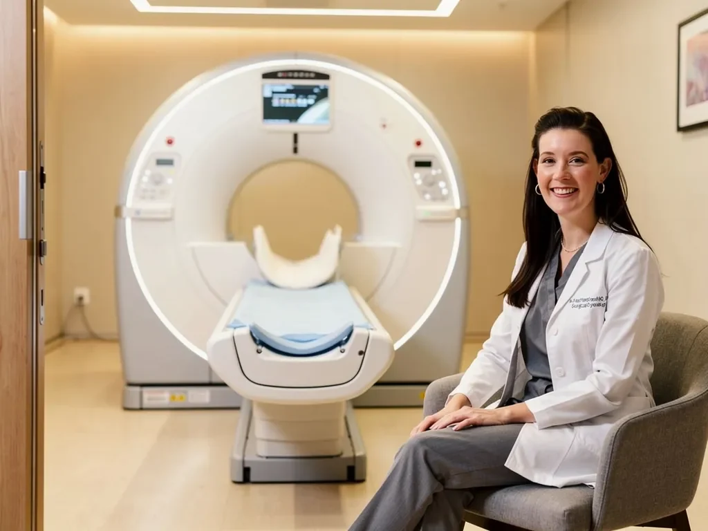
Knowing what the examination is intended to answer can make the imaging process more useful and reduce uncertainty when the report becomes available.
Questions Before Scheduling the MRI
Patients may ask:
-
Is this a dedicated pelvic MRI for endometriosis?
-
Will it assess the bowel, bladder, ureters, uterosacral ligaments, and ovaries when relevant?
-
Does the imaging center regularly perform endometriosis studies?
-
Will a radiologist experienced in gynecologic imaging interpret it?
-
Is intravenous contrast expected?
-
What preparation is required?
-
Should I bring previous ultrasound images, MRI studies, or operative reports?
-
Are there any MRI safety concerns I should discuss in advance?
Questions After Receiving the Report
Useful questions include:
-
Did the MRI identify a definite lesion or only indirect signs?
-
Which pelvic structures appear involved?
-
Were the bowel, bladder, ureters, ovaries, and uterosacral ligaments assessed?
-
Does the report suggest an endometrioma, adenomyosis, or another condition?
-
What types of endometriosis does this scan not rule out?
-
Would specialist ultrasound add useful information?
-
Would specialist review of the actual images change the interpretation?
-
What is the next step if my symptoms continue?
Questions When Surgery Is Being Discussed
Patients may ask:
-
How do the MRI findings affect the proposed procedure?
-
Is bowel, bladder, ureteral, or multidisciplinary surgical involvement anticipated?
-
Which findings are definite and which remain uncertain?
-
Are nonsurgical treatment options still reasonable?
-
How do my symptoms and goals affect the recommendation?
-
Could additional findings be discovered during surgery?
-
What symptoms may have causes that surgery will not address?
Frequently Asked Questions
Can an MRI Detect Deep Endometriosis?
Yes. A dedicated pelvic MRI can identify many forms of deep endometriosis and help map their relationship to surrounding organs.
Its performance varies according to lesion location, protocol, technical quality, disease characteristics, and radiologist expertise. MRI is useful but not infallible.
Can a Normal MRI Rule Out Endometriosis?
No. A normal MRI does not exclude superficial peritoneal endometriosis, every small deep lesion, or other causes of pelvic pain.
Current guidance states that endometriosis should not be excluded solely because examination or imaging is normal.
Is MRI Better Than Transvaginal Ultrasound?
Not in every situation.
Specialist ultrasound can detect endometriomas and many forms of deep endometriosis while providing dynamic information about organ mobility and tenderness. MRI provides a broad pelvic overview and may be particularly useful for complex, multifocal, or difficult-to-map disease.
The better examination depends on the suspected location, patient tolerance, available expertise, and treatment-planning needs.
Can MRI Detect Bowel Endometriosis?
MRI can identify many rectal and sigmoid lesions and help estimate their location, length, depth, and relationship to surrounding tissues.
It cannot determine every microscopic detail, and specialist ultrasound, additional imaging, or surgical evaluation may still be needed.
Can MRI Detect Bladder or Ureteral Endometriosis?
MRI may identify deep lesions, fibrosis, anatomical distortion, or urinary tract involvement affecting the bladder or ureters.
When obstruction or ureteral disease is suspected, additional urinary tract assessment and specialist coordination may be required.
Can MRI Detect Superficial Endometriosis?
MRI is less reliable for small or superficial peritoneal lesions.
A negative scan should not be used to exclude superficial disease when the symptom history remains suggestive.
Does an Endometriosis MRI Require Contrast?
Not always.
Many characteristic findings are assessed using noncontrast T1- and T2-weighted imaging. Contrast may be used when an ovarian mass is indeterminate or another diagnostic question requires further characterization.
Does a Positive MRI Mean I Need Surgery?
No.
Treatment depends on symptoms, organ function, previous treatment, fertility goals, risks, and patient preferences. Imaging informs the decision but does not make it automatically.
Is Laparoscopy Still Necessary After MRI?
Not for every patient.
Clinical evaluation and imaging may support diagnosis and allow treatment to begin without surgery. Laparoscopy may be considered when symptoms remain severe, treatment has been unsuccessful, imaging suggests disease requiring surgery, or diagnostic and surgical evaluation is otherwise appropriate.
Should an Endometriosis Specialist Review My MRI?
Specialist review may be particularly useful when:
-
The report is inconclusive
-
Symptoms and imaging do not match
-
Deep or multiorgan disease is suspected
-
Previous surgery has altered the anatomy
-
Surgery is being planned
The specialist should review the MRI within the context of the patient’s symptoms, examination, previous treatment, and goals.
Conclusion
A pelvic MRI can be a valuable tool for detecting and mapping deep endometriosis, particularly when disease may involve the bowel, bladder, ureters, ovaries, uterosacral ligaments, rectovaginal region, or several pelvic compartments.
Its value depends on the clinical question, imaging protocol, technical quality, and experience of the interpreting radiologist. A dedicated endometriosis MRI can provide much more useful information than a general pelvic study when complex disease is suspected.
Patients should remember three central points:
-
MRI is most useful for mapping many forms of deep or complex disease, not for ruling out every form of endometriosis.
-
A normal MRI does not end the diagnostic process when symptoms remain persistent or disabling.
-
Imaging should guide individualized treatment planning without automatically committing a patient to surgery.
At the Endometriosis Center of Excellence, Dr. Rachael Haverland evaluates imaging alongside pelvic pain, menstrual symptoms, bowel and bladder concerns, previous treatment, surgical history, fertility goals, and daily functional impact. This comprehensive assessment helps determine whether the most appropriate next step involves specialist image review, medical management, pelvic floor care, minimally invasive excision surgery, or coordination with additional specialists.